6BEA | pdb_00006bea

Crystal structure of the autotransporter UpaB from E. coli strain CFT073


Experimental Data Snapshot

  • Method: X-RAY DIFFRACTION
  • Resolution: 1.97 Å
  • R-Value Free: 
    0.218 (Depositor), 0.219 (DCC) 
  • R-Value Work: 
    0.175 (Depositor), 0.175 (DCC) 
  • R-Value Observed: 
    0.177 (Depositor) 

wwPDB Validation 3D Report Full Report

Validation slider image for 6BEA

This is version 1.4 of the entry. See complete history

Literature

Unique structural features of a bacterial autotransporter adhesin suggest mechanisms for interaction with host macromolecules.

Paxman, J.J.Lo, A.W.Sullivan, M.J.Panjikar, S.Kuiper, M.Whitten, A.E.Wang, G.Luan, C.H.Moriel, D.G.Tan, L.Peters, K.M.Phan, M.D.Gee, C.L.Ulett, G.C.Schembri, M.A.Heras, B.

(2019) Nat Commun 10: 1967-1967

  • DOI: https://doi.org/10.1038/s41467-019-09814-6
  • Primary Citation Related Structures: 
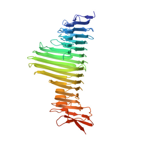
    6BEA

  • PubMed Abstract: 

    Autotransporters are the largest family of outer membrane and secreted proteins in Gram-negative bacteria. Most autotransporters are localised to the bacterial surface where they promote colonisation of host epithelial surfaces. Here we present the crystal structure of UpaB, an autotransporter that is known to contribute to uropathogenic E. coli (UPEC) colonisation of the urinary tract. We provide evidence that UpaB can interact with glycosaminoglycans and host fibronectin. Unique modifications to its core β-helical structure create a groove on one side of the protein for interaction with glycosaminoglycans, while the opposite face can bind fibronectin. Our findings reveal far greater diversity in the autotransporter β-helix than previously thought, and suggest that this domain can interact with host macromolecules. The relevance of these interactions during infection remains unclear.


  • Organizational Affiliation
    • Department of Biochemistry and Genetics, La Trobe Institute for Molecular Science, La Trobe University, Melbourne, 3086, VIC, Australia.

Macromolecule Content 

  • Total Structure Weight: 48.67 kDa 
  • Atom Count: 3,443 
  • Modeled Residue Count: 433 
  • Deposited Residue Count: 466 
  • Unique protein chains: 1

Macromolecules

Find similar proteins by:|  3D Structure
Entity ID: 1
MoleculeChains  Sequence LengthOrganismDetailsImage
Autotransporter protein UpaB466Escherichia coli CFT073Mutation(s): 0 
Gene Names: c0426
Membrane Entity: Yes 
UniProt
Find proteins for A0A0H2V5A3 (Escherichia coli O6:H1 (strain CFT073 / ATCC 700928 / UPEC))
Explore A0A0H2V5A3 
Go to UniProtKB:  A0A0H2V5A3
Entity Groups
Sequence Clusters30% Identity50% Identity70% Identity90% Identity95% Identity100% Identity
UniProt GroupA0A0H2V5A3
Sequence Annotations
Expand
Reference Sequence

Small Molecules

Ligands 6 Unique
IDChains Name / Formula / InChI Key2D Diagram3D Interactions
P6G

Query on P6G


Download:Ideal Coordinates CCD File
F [auth A]HEXAETHYLENE GLYCOL
C12 H26 O7
IIRDTKBZINWQAW-UHFFFAOYSA-N
SO4

Query on SO4


Download:Ideal Coordinates CCD File
G [auth A]SULFATE ION
O4 S
QAOWNCQODCNURD-UHFFFAOYSA-L
GOL

Query on GOL


Download:Ideal Coordinates CCD File
B [auth A],
C [auth A],
D [auth A],
E [auth A],
K [auth A]
GLYCEROL
C3 H8 O3
PEDCQBHIVMGVHV-UHFFFAOYSA-N
CA

Query on CA


Download:Ideal Coordinates CCD File
H [auth A]CALCIUM ION
Ca
BHPQYMZQTOCNFJ-UHFFFAOYSA-N
CL

Query on CL


Download:Ideal Coordinates CCD File
I [auth A]CHLORIDE ION
Cl
VEXZGXHMUGYJMC-UHFFFAOYSA-M
NA

Query on NA


Download:Ideal Coordinates CCD File
J [auth A]SODIUM ION
Na
FKNQFGJONOIPTF-UHFFFAOYSA-N

Experimental Data & Validation

Experimental Data

  • Method: X-RAY DIFFRACTION
  • Resolution: 1.97 Å
  • R-Value Free:  0.218 (Depositor), 0.219 (DCC) 
  • R-Value Work:  0.175 (Depositor), 0.175 (DCC) 
  • R-Value Observed: 0.177 (Depositor) 
Space Group: P 31 2 1
Unit Cell:
Length ( Å )Angle ( ˚ )
a = 68.611α = 90
b = 68.611β = 90
c = 165.568γ = 120
Software Package:
Software NamePurpose
PHENIXrefinement
HKL-2000data scaling
PDB_EXTRACTdata extraction
SHELXDEphasing
ARP/wARPmodel building
Cootmodel building
HKL-2000data reduction

Structure Validation

View Full Validation Report


Entry History 

& Funding Information

Deposition Data


Funding OrganizationLocationGrant Number
Australian Research Council (ARC)AustraliaDP150102287
Australian Research Council (ARC)AustraliaFT130100580
Australian Synchrotron Research Program FellowshipAustralia--

Revision History  (Full details and data files)

  • Version 1.0: 2019-04-10
    Type: Initial release
  • Version 1.1: 2019-05-08
    Changes: Data collection, Database references
  • Version 1.2: 2019-05-15
    Changes: Data collection, Database references
  • Version 1.3: 2020-01-01
    Changes: Author supporting evidence
  • Version 1.4: 2024-03-13
    Changes: Data collection, Database references