RCSB PDB - 6BDK: Crystal structure of human CYP3A4 bound to an inhibitor

 6BDK

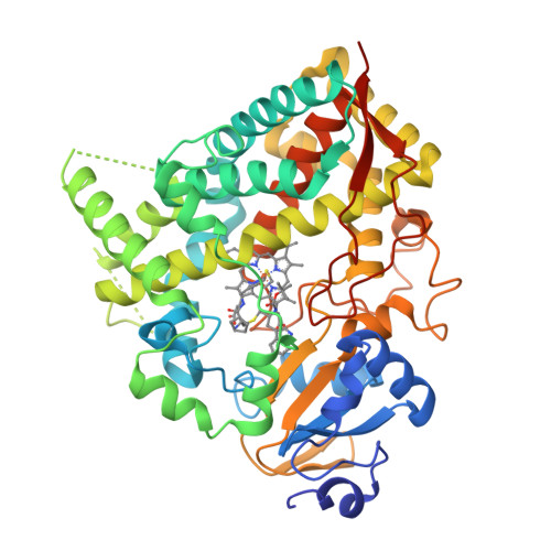
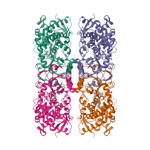
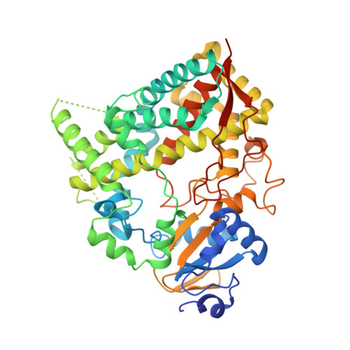
Crystal structure of human CYP3A4 bound to an inhibitor


Experimental Data Snapshot

  • Method: X-RAY DIFFRACTION
  • Resolution: 2.67 Å
  • R-Value Free: 0.272 
  • R-Value Work: 0.205 
  • R-Value Observed: 0.209 

Starting Model: experimental
View more details

wwPDB Validation   3D Report Full Report


Ligand Structure Quality Assessment 

Created with Raphaël 2.3.0Worse 01 BetterLigand structure goodness of fit to experimental dataBest fitted HEMClick on this verticalbar to view detailsBest fitted DJ1Click on this verticalbar to view details

This is version 1.4 of the entry. See complete history


Literature

Inhibition of Human CYP3A4 by Rationally Designed Ritonavir-Like Compounds: Impact and Interplay of the Side Group Functionalities.

Samuels, E.R.Sevrioukova, I.

(2018) Mol Pharm 15: 279-288

  • DOI: https://doi.org/10.1021/acs.molpharmaceut.7b00957
  • Primary Citation of Related Structures:  
    6BCZ, 6BD5, 6BD6, 6BDK

  • PubMed Abstract: 

    Structure-function relationships of nine rationally designed ritonavir-like compounds were investigated to better understand the ligand binding and inhibitory mechanism in human drug-metabolizing cytochrome P450 3A4 (CYP3A4). The analogs had a similar backbone and pyridine and tert-butyloxycarbonyl (Boc) as the heme-ligating and terminal groups, respectively. N-Isopropyl, N-cyclopentyl, or N-phenyl were the R 1 -side group substituents alone (compounds 5a-c) or in combination with phenyl or indole at the R 2 position (8a-c and 8d-f subseries, respectively). Our experimental and structural data indicate that (i) for all analogs, a decrease in the dissociation constant (K s ) coincides with a decrease in IC 50 , but no relation with other derived parameters is observed; (ii) an increase in the R 1 volume, hydrophobicity, and aromaticity markedly lowers K s and IC 50 , whereas the addition of aromatic R 2 has a more pronounced positive effect on the inhibitory potency than the binding strength; (iii) the ligands' association mode is strongly influenced by the mutually dependent R 1 -R 2 interplay, but the R 1 -mediated interactions are dominant and define the overall conformation in the active site; (iv) formation of a strong H-bond with Ser119 is a prerequisite for potent CYP3A4 inhibition; and (v) the strongest inhibitor in the series, the R 1 -phenyl/R 2 -indole containing 8f (K s and IC 50 of 0.08 and 0.43 μM, respectively), is still less potent than ritonavir, even under conditions that prevent the mechanism based inactivation of CYP3A4. Crystallographic data were essential for better understanding and interpretation of the experimental results, and suggested how the inhibitor design could be further optimized.


  • Organizational Affiliation

    Departments of Pharmaceutical Sciences and ‡Molecular Biology and Biochemistry, University of California , Irvine, California 92697-3900, United States.


Macromolecules
Find similar proteins by:  (by identity cutoff)  |  3D Structure
Entity ID: 1
MoleculeChains Sequence LengthOrganismDetailsImage
Cytochrome P450 3A4487Homo sapiensMutation(s): 0 
Gene Names: CYP3A4CYP3A3
EC: 1.14.13 (PDB Primary Data), 1.14.13.157 (PDB Primary Data), 1.14.13.32 (PDB Primary Data), 1.14.14.1 (PDB Primary Data), 1.14.13.67 (PDB Primary Data), 1.14.13.97 (PDB Primary Data), 1.14.14.73 (UniProt), 1.14.14.56 (UniProt), 1.14.14.55 (UniProt)
UniProt & NIH Common Fund Data Resources
Find proteins for P08684 (Homo sapiens)
Explore P08684 
Go to UniProtKB:  P08684
PHAROS:  P08684
GTEx:  ENSG00000160868 
Entity Groups  
Sequence Clusters30% Identity50% Identity70% Identity90% Identity95% Identity100% Identity
UniProt GroupP08684
Sequence Annotations
Expand
  • Reference Sequence
Small Molecules
Ligands 2 Unique
IDChains Name / Formula / InChI Key2D Diagram3D Interactions
HEM
Query on HEM

Download Ideal Coordinates CCD File 
B [auth A]PROTOPORPHYRIN IX CONTAINING FE
C34 H32 Fe N4 O4
KABFMIBPWCXCRK-RGGAHWMASA-L
DJ1
Query on DJ1

Download Ideal Coordinates CCD File 
C [auth A]tert-butyl [(2S)-1-{[(2R)-2-(cyclopentylamino)-3-oxo-3-{[(pyridin-3-yl)methyl]amino}propyl]sulfanyl}-3-(1H-indol-3-yl)propan-2-yl]carbamate
C30 H41 N5 O3 S
AQDOXNFDADPPBW-IGKIAQTJSA-N
Experimental Data & Validation

Experimental Data

  • Method: X-RAY DIFFRACTION
  • Resolution: 2.67 Å
  • R-Value Free: 0.272 
  • R-Value Work: 0.205 
  • R-Value Observed: 0.209 
  • Space Group: I 2 2 2
Unit Cell:
Length ( Å )Angle ( ˚ )
a = 77.479α = 90
b = 102.39β = 90
c = 127.76γ = 90
Software Package:
Software NamePurpose
PHENIXrefinement
MOSFLMdata reduction
SCALAdata scaling
PHASERphasing

Structure Validation

View Full Validation Report


Ligand Structure Quality Assessment 

Created with Raphaël 2.3.0Worse 01 BetterLigand structure goodness of fit to experimental dataBest fitted HEMClick on this verticalbar to view detailsBest fitted DJ1Click on this verticalbar to view details

Entry History & Funding Information

Deposition Data


Funding OrganizationLocationGrant Number
National Institutes of Health/National Institute of Environmental Health Sciences (NIH/NIEHS)United StatesES025767

Revision History  (Full details and data files)

  • Version 1.0: 2017-12-13
    Type: Initial release
  • Version 1.1: 2018-01-17
    Changes: Author supporting evidence
  • Version 1.2: 2019-12-18
    Changes: Author supporting evidence
  • Version 1.3: 2020-01-29
    Changes: Database references, Derived calculations
  • Version 1.4: 2023-10-04
    Changes: Data collection, Database references, Refinement description