6B6I | pdb_00006b6i

2.4A resolution structure of human Norovirus GII.4 protease


Experimental Data Snapshot

  • Method: X-RAY DIFFRACTION
  • Resolution: 2.44 Å
  • R-Value Free: 
    0.222 (Depositor), 0.228 (DCC) 
  • R-Value Work: 
    0.180 (Depositor), 0.190 (DCC) 
  • R-Value Observed: 
    0.182 (Depositor) 

Starting Model: experimental
View more details

wwPDB Validation 3D Report Full Report

Validation slider image for 6B6I

This is version 1.3 of the entry. See complete history

Literature

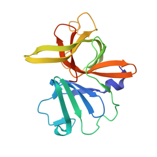
Structural and Antiviral Studies of the Human Norovirus GII.4 Protease.

Muzzarelli, K.M.Kuiper, B.Spellmon, N.Brunzelle, J.Hackett, J.Amblard, F.Zhou, S.Liu, P.Kovari, I.A.Yang, Z.Schinazi, R.F.Kovari, L.C.

(2019) Biochemistry 58: 900-907

  • DOI: https://doi.org/10.1021/acs.biochem.8b01063
  • Primary Citation Related Structures: 
    6B6I

  • PubMed Abstract: 

    Norovirus is the leading cause of acute gastroenteritis worldwide with a yearly reported 700 million cases driving a $60 billion global socioeconomic burden. With no United States Food and Drug Administration approved therapeutics and the chance for severe chronic infection and life-threatening complications, researchers have identified the protease as a potential target. However, drug development has focused on the norovirus GI.1 strain despite its accounting for less than 5% of all outbreaks. Our lab aims to change focus for norovirus drug design from GI.1 to the highly infective GII.4, responsible for more than 50% of all outbreaks worldwide. With the first published crystal structure of the norovirus GII.4 protease, we have identified several significant differences in the structure and active site that have hindered development of a potent inhibitor targeting the norovirus GII.4 protease. With these new insights, we have begun designing compounds that demonstrate increased inhibition of the clinically most relevant norovirus GII.4 strain.


  • Organizational Affiliation
    • Department of Biochemistry, Microbiology and Immunology , Wayne State University School of Medicine , Detroit , Michigan 48201 , United States.

Macromolecule Content 

  • Total Structure Weight: 153.94 kDa 
  • Atom Count: 10,640 
  • Modeled Residue Count: 1,409 
  • Deposited Residue Count: 1,448 
  • Unique protein chains: 1

Macromolecules

Find similar proteins by:|  3D Structure
Entity ID: 1
MoleculeChains  Sequence LengthOrganismDetailsImage
3C-like protease
A, B, C, D, E
A, B, C, D, E, F, G, H
181Norovirus GII.4Mutation(s): 1 
UniProt
Find proteins for A0A0K2E2J2 (Norovirus Hu/GII.P4_GII.4/Minerva/2006/USA)
Explore A0A0K2E2J2 
Go to UniProtKB:  A0A0K2E2J2
Entity Groups
Sequence Clusters30% Identity50% Identity70% Identity90% Identity95% Identity100% Identity
UniProt GroupA0A0K2E2J2
Sequence Annotations
Expand
Reference Sequence

Experimental Data & Validation

Experimental Data

  • Method: X-RAY DIFFRACTION
  • Resolution: 2.44 Å
  • R-Value Free:  0.222 (Depositor), 0.228 (DCC) 
  • R-Value Work:  0.180 (Depositor), 0.190 (DCC) 
  • R-Value Observed: 0.182 (Depositor) 
Space Group: P 32
Unit Cell:
Length ( Å )Angle ( ˚ )
a = 112.94α = 90
b = 112.94β = 90
c = 153.37γ = 120
Software Package:
Software NamePurpose
PHENIXrefinement
XDSdata reduction
Aimlessdata scaling
PHENIXphasing

Structure Validation

View Full Validation Report


Entry History 

& Funding Information

Deposition Data


Funding OrganizationLocationGrant Number
Wayne State UniversityUnited States--

Revision History  (Full details and data files)

  • Version 1.0: 2018-10-03
    Type: Initial release
  • Version 1.1: 2019-01-16
    Changes: Data collection, Database references
  • Version 1.2: 2019-02-27
    Changes: Data collection, Database references
  • Version 1.3: 2023-10-04
    Changes: Data collection, Database references, Refinement description