6B09 | pdb_00006b09

Crystal structure of HsNUDT16 in complex with diADPR (soaked)


Experimental Data Snapshot

  • Method: X-RAY DIFFRACTION
  • Resolution: 3.20 Å
  • R-Value Free: 
    0.279 (Depositor), 0.278 (DCC) 
  • R-Value Work: 
    0.256 (Depositor), 0.256 (DCC) 
  • R-Value Observed: 
    0.257 (Depositor) 

wwPDB Validation 3D Report Full Report

Validation slider image for 6B09

Ligand Structure Quality Assessment 


This is version 1.2 of the entry. See complete history

Literature

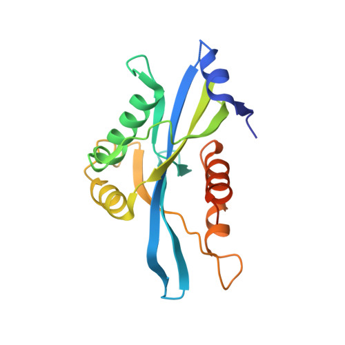
Structural analyses of NudT16-ADP-ribose complexes direct rational design of mutants with improved processing of poly(ADP-ribosyl)ated proteins.

Thirawatananond, P.McPherson, R.L.Malhi, J.Nathan, S.Lambrecht, M.J.Brichacek, M.Hergenrother, P.J.Leung, A.K.L.Gabelli, S.B.

(2019) Sci Rep 9: 5940-5940

  • DOI: https://doi.org/10.1038/s41598-019-39491-w
  • Primary Citation Related Structures: 
    5VY2, 5W6X, 5W6Z, 5WJI, 6B09

  • PubMed Abstract: 

    ADP-ribosylation is a post-translational modification that occurs on chemically diverse amino acids, including aspartate, glutamate, lysine, arginine, serine and cysteine on proteins and is mediated by ADP-ribosyltransferases, including a subset commonly known as poly(ADP-ribose) polymerases. ADP-ribose can be conjugated to proteins singly as a monomer or in polymeric chains as poly(ADP-ribose). While ADP-ribosylation can be reversed by ADP-ribosylhydrolases, this protein modification can also be processed to phosphoribosylation by enzymes possessing phosphodiesterase activity, such as snake venom phosphodiesterase, mammalian ectonucleotide pyrophosphatase/phosphodiesterase 1, Escherichia coli RppH, Legionella pneumophila Sde and Homo sapiens NudT16 (HsNudT16). Our studies here sought to utilize X-ray crystallographic structures of HsNudT16 in complex with monomeric and dimeric ADP-ribose in identifying the active site for binding and processing free and protein-conjugated ADP-ribose into phosphoribose forms. These structural data guide rational design of mutants that widen the active site to better accommodate protein-conjugated ADP-ribose. We identified that several HsNudT16 mutants (Δ17, F36A, and F61S) have reduced activity for free ADP-ribose, similar processing ability against protein-conjugated mono(ADP-ribose), but improved catalytic efficiency for protein-conjugated poly(ADP-ribose). These HsNudT16 variants may, therefore, provide a novel tool to investigate different forms of ADP-ribose.


  • Organizational Affiliation
    • Department of Biophysics and Biophysical Chemistry, Johns Hopkins University School of Medicine, Baltimore, MD, 21205, USA.

Macromolecule Content 

  • Total Structure Weight: 44.78 kDa 
  • Atom Count: 2,874 
  • Modeled Residue Count: 351 
  • Deposited Residue Count: 390 
  • Unique protein chains: 1

Macromolecules

Find similar proteins by:|  3D Structure
Entity ID: 1
MoleculeChains  Sequence LengthOrganismDetailsImage
U8 snoRNA-decapping enzyme
A, B
195Homo sapiensMutation(s): 1 
Gene Names: NUDT16
EC: 3.6.1.62 (PDB Primary Data), 3.6.1.64 (PDB Primary Data)
UniProt & NIH Common Fund Data Resources
Find proteins for Q96DE0 (Homo sapiens)
Explore Q96DE0 
Go to UniProtKB:  Q96DE0
PHAROS:  Q96DE0
GTEx:  ENSG00000198585 
Entity Groups
Sequence Clusters30% Identity50% Identity70% Identity90% Identity95% Identity100% Identity
UniProt GroupQ96DE0
Sequence Annotations
Expand
Reference Sequence

Small Molecules

Ligands 4 Unique
IDChains Name / Formula / InChI Key2D Diagram3D Interactions
C7G

Query on C7G


Download:Ideal Coordinates CCD File
C [auth A],
H [auth B]
[(2~{R},3~{S},4~{S},5~{S})-5-(6-aminopurin-9-yl)-4-[(2~{S},3~{S},4~{S},5~{S})-5-[[[[(2~{R},3~{R},4~{S},5~{S})-5-(6-aminopurin-9-yl)-3,4-bis(oxidanyl)oxolan-2-yl]methoxy-oxidanyl-phosphoryl]oxy-oxidanyl-phosphoryl]oxymethyl]-3,4-bis(oxidanyl)oxolan-2-yl]oxy-3-oxidanyl-oxolan-2-yl]methyl phosphono hydrogen phosphate
C25 H36 N10 O23 P4
CSEMRHUJDKFXPV-OATIKAJMSA-N
CL

Query on CL


Download:Ideal Coordinates CCD File
F [auth A]CHLORIDE ION
Cl
VEXZGXHMUGYJMC-UHFFFAOYSA-M
MG

Query on MG


Download:Ideal Coordinates CCD File
D [auth A],
E [auth A],
I [auth B],
J [auth B]
MAGNESIUM ION
Mg
JLVVSXFLKOJNIY-UHFFFAOYSA-N
NA

Query on NA


Download:Ideal Coordinates CCD File
G [auth A],
K [auth B]
SODIUM ION
Na
FKNQFGJONOIPTF-UHFFFAOYSA-N

Experimental Data & Validation

Experimental Data

  • Method: X-RAY DIFFRACTION
  • Resolution: 3.20 Å
  • R-Value Free:  0.279 (Depositor), 0.278 (DCC) 
  • R-Value Work:  0.256 (Depositor), 0.256 (DCC) 
  • R-Value Observed: 0.257 (Depositor) 
Space Group: C 1 2 1
Unit Cell:
Length ( Å )Angle ( ˚ )
a = 113.395α = 90
b = 47.063β = 108.74
c = 75.792γ = 90
Software Package:
Software NamePurpose
REFMACrefinement
StructureStudiodata collection
SCALEPACKdata scaling
PDB_EXTRACTdata extraction
Cootmodel building
HKL-3000data reduction
MOLREPphasing

Structure Validation

View Full Validation Report


Ligand Structure Quality Assessment 


Entry History 

Deposition Data

Revision History  (Full details and data files)

  • Version 1.0: 2019-01-09
    Type: Initial release
  • Version 1.1: 2019-04-24
    Changes: Data collection, Database references
  • Version 1.2: 2024-03-13
    Changes: Data collection, Database references, Derived calculations