6B07 | pdb_00006b07

Crystal structure of CfFPPS2, a lepidopteran type-II farnesyl diphosphate synthase, complexed with [1-phosphono-2-(1-propylpyridin-2-yl)ethyl]phosphonic acid (inhibitor 1d)


Experimental Data Snapshot

  • Method: X-RAY DIFFRACTION
  • Resolution: 1.98 Å
  • R-Value Free: 
    0.209 (Depositor), 0.215 (DCC) 
  • R-Value Work: 
    0.171 (Depositor), 0.180 (DCC) 
  • R-Value Observed: 
    0.173 (Depositor) 

Starting Model: experimental
View more details

wwPDB Validation 3D Report Full Report

Validation slider image for 6B07

Ligand Structure Quality Assessment 


This is version 2.0 of the entry. See complete history

Literature

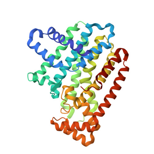
Structural characterization of a lepidopteran type-II farnesyl diphosphate synthase from the spruce budworm, Choristoneura fumiferana: Implications for inhibitor design.

Picard, M.E.Nisole, A.Beliveau, C.Sen, S.Barbar, A.Shi, R.Cusson, M.

(2017) Insect Biochem Mol Biol 92: 84-92

  • DOI: https://doi.org/10.1016/j.ibmb.2017.11.011
  • Primary Citation Related Structures: 
    6B02, 6B04, 6B06, 6B07

  • PubMed Abstract: 

    Farnesyl diphosphate synthase (FPPS) is an enzyme from the class of short chain (E)-prenyltransferases that catalyzes the condensation of two molecules of isopentenyl diphosphate (IPP, C 5 ) with dimethylallyl diphosphate (DMAPP, C 5 ) to generate the C 15 product FPP. In insects, FPPS plays a key role in the biosynthesis of the morphogenetic and gonadotropic "juvenile hormone" (JH). Lepidopteran genomes encode two very distinct FPPS paralogs, one of which ("type-II") is expressed almost exclusively in the JH-producing glands, the corpora allata. This paralog has been hypothesized to display structural features that enable the binding of the bulkier precursors required for the biosynthesis of lepidopteran ethyl-branched JHs. Here, we report on the first crystal structures of an insect FPPS solved to date. Apo, ligand-bound, and inhibitor-bound structures of type-II FPPS (FPPS2) from the spruce budworm, Choristoneura fumiferana (Order: Lepidoptera), were obtained. Comparison of apo and inhibitor-bound enzymes revealed differences in both inhibitor binding and structural plasticity of CfFPPS2 compared to other FPPSs. Our data showed that IPP is not essential to the closure of the C-terminal tail. Ortho-substituted pyridinium bisphosphonates, previously shown to inhibit CfFPPS2, bound to the allylic site, as predicted; however, their alkyl groups were oriented towards the homoallylic binding site, with the bulkier propyl-substituted inhibitor penetrating deeply into the IPP binding pocket. The current study sheds light on the structural basis of substrate specificity of type-II FPPS of the spruce budworm. Through a comparison with other inhibitor-bound FPPSs, we propose several approaches to improve inhibitor selectivity and potency.


  • Organizational Affiliation
    • Département de biochimie, de microbiologie et de bio-informatique, Institut de Biologie Intégrative et des Systèmes, PROTEO, Université Laval, Quebec City, QC, G1V 0A6, Canada. Electronic address: marie-eve.picard.8@ulaval.ca.

Macromolecule Content 

  • Total Structure Weight: 119.07 kDa 
  • Atom Count: 9,174 
  • Modeled Residue Count: 1,013 
  • Deposited Residue Count: 1,023 
  • Unique protein chains: 1

Macromolecules

Find similar proteins by:|  3D Structure
Entity ID: 1
MoleculeChains  Sequence LengthOrganismDetailsImage
Farnesyl diphosphate synthase
A, B, C
341Choristoneura fumiferanaMutation(s): 0 
EC: 2.5.1
UniProt
Find proteins for Q1XAB1 (Choristoneura fumiferana)
Explore Q1XAB1 
Go to UniProtKB:  Q1XAB1
Entity Groups
Sequence Clusters30% Identity50% Identity70% Identity90% Identity95% Identity100% Identity
UniProt GroupQ1XAB1
Sequence Annotations
Expand
Reference Sequence

Small Molecules

Ligands 3 Unique
IDChains Name / Formula / InChI Key2D Diagram3D Interactions
C6M

Query on C6M


Download:Ideal Coordinates CCD File
D [auth A],
J [auth B],
P [auth C]
2-(2,2-diphosphonoethyl)-1-propylpyridin-1-ium
C10 H18 N O6 P2
KNULKELAVSFYBS-UHFFFAOYSA-O
EDO

Query on EDO


Download:Ideal Coordinates CCD File
H [auth A]
I [auth A]
N [auth B]
O [auth B]
T [auth C]
H [auth A],
I [auth A],
N [auth B],
O [auth B],
T [auth C],
U [auth C],
V [auth C]
1,2-ETHANEDIOL
C2 H6 O2
LYCAIKOWRPUZTN-UHFFFAOYSA-N
MG

Query on MG


Download:Ideal Coordinates CCD File
E [auth A]
F [auth A]
G [auth A]
K [auth B]
L [auth B]
E [auth A],
F [auth A],
G [auth A],
K [auth B],
L [auth B],
M [auth B],
Q [auth C],
R [auth C],
S [auth C]
MAGNESIUM ION
Mg
JLVVSXFLKOJNIY-UHFFFAOYSA-N

Experimental Data & Validation

Experimental Data

  • Method: X-RAY DIFFRACTION
  • Resolution: 1.98 Å
  • R-Value Free:  0.209 (Depositor), 0.215 (DCC) 
  • R-Value Work:  0.171 (Depositor), 0.180 (DCC) 
  • R-Value Observed: 0.173 (Depositor) 
Space Group: C 1 2 1
Unit Cell:
Length ( Å )Angle ( ˚ )
a = 186.913α = 90
b = 122.579β = 106.51
c = 69.064γ = 90
Software Package:
Software NamePurpose
REFMACrefinement
SCALEPACKdata scaling
PDB_EXTRACTdata extraction
DENZOdata reduction
MOLREPphasing

Structure Validation

View Full Validation Report


Ligand Structure Quality Assessment 


Entry History 

& Funding Information

Deposition Data


Funding OrganizationLocationGrant Number
Natural Sciences and Engineering Research Council (NSERC, Canada)Canada436202

Revision History  (Full details and data files)

  • Version 1.0: 2017-12-13
    Type: Initial release
  • Version 1.1: 2018-01-10
    Changes: Database references
  • Version 1.2: 2018-01-17
    Changes: Author supporting evidence
  • Version 1.3: 2020-01-08
    Changes: Author supporting evidence
  • Version 1.4: 2023-10-04
    Changes: Data collection, Database references, Derived calculations, Refinement description
  • Version 2.0: 2024-05-01
    Changes: Derived calculations, Non-polymer description, Structure summary