6AUE | pdb_00006aue

Artificial Metalloproteins Containing a Co4O4 Active Site - 2xm-S112Y-b


Experimental Data Snapshot

  • Method: X-RAY DIFFRACTION
  • Resolution: 1.36 Å
  • R-Value Free: 
    0.229 (Depositor), 0.230 (DCC) 
  • R-Value Work: 
    0.203 (Depositor), 0.206 (DCC) 
  • R-Value Observed: 
    0.204 (Depositor) 

Starting Model: experimental
View more details

wwPDB Validation 3D Report Full Report

Validation slider image for 6AUE

Ligand Structure Quality Assessment 


This is version 1.4 of the entry. See complete history

Literature

Artificial Metalloproteins Containing Co

Olshansky, L.Huerta-Lavorie, R.Nguyen, A.I.Vallapurackal, J.Furst, A.Tilley, T.D.Borovik, A.S.

(2018) J Am Chem Soc 140: 2739-2742

  • DOI: https://doi.org/10.1021/jacs.7b13052
  • Primary Citation Related Structures: 
    6AUC, 6AUE, 6AUH, 6AUL, 6AUO

  • PubMed Abstract: 

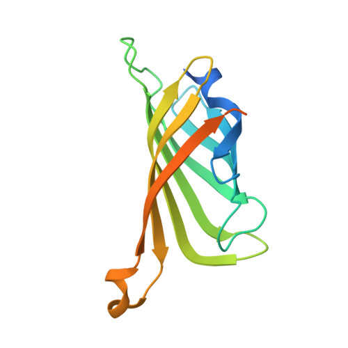
    Artificial metalloproteins (ArMs) containing Co 4 O 4 cubane active sites were constructed via biotin-streptavidin technology. Stabilized by hydrogen bonds (H-bonds), terminal and cofacial Co III -OH 2 moieties are observed crystallographically in a series of immobilized cubane sites. Solution electrochemistry provided correlations of oxidation potential and pH. For variants containing Ser and Phe adjacent to the metallocofactor, 1e - /1H + chemistry predominates until pH 8, above which the oxidation becomes pH-independent. Installation of Tyr proximal to the Co 4 O 4 active site provided a single H-bond to one of a set of cofacial Co III -OH 2 groups. With this variant, multi-e - /multi-H + chemistry is observed, along with a change in mechanism at pH 9.5 that is consistent with Tyr deprotonation. With structural similarities to both the oxygen-evolving complex of photosystem II (H-bonded Tyr) and to thin film water oxidation catalysts (Co 4 O 4 core), these findings bridge synthetic and biological systems for water oxidation, highlighting the importance of secondary sphere interactions in mediating multi-e - /multi-H + reactivity.


  • Organizational Affiliation
    • Department of Chemistry, University of California, Irvine , Irvine, California 92697, United States.

Macromolecule Content 

  • Total Structure Weight: 17.63 kDa 
  • Atom Count: 1,050 
  • Modeled Residue Count: 121 
  • Deposited Residue Count: 159 
  • Unique protein chains: 1

Macromolecules

Find similar proteins by:|  3D Structure
Entity ID: 1
MoleculeChains  Sequence LengthOrganismDetailsImage
Streptavidin159Streptomyces avidiniiMutation(s): 3 
UniProt
Find proteins for P22629 (Streptomyces avidinii)
Explore P22629 
Go to UniProtKB:  P22629
Entity Groups
Sequence Clusters30% Identity50% Identity70% Identity90% Identity95% Identity100% Identity
UniProt GroupP22629
Sequence Annotations
Expand
Reference Sequence

Small Molecules

Ligands 1 Unique
IDChains Name / Formula / InChI Key2D Diagram3D Interactions
OL3

Query on OL3


Download:Ideal Coordinates CCD File
B [auth A]N-biotin-C-Co4(mu3-O)4(OAc)(Py)4(H2O)3-beta-alanine
C35 H45 Co4 N7 O13 S
JXTIQCHHLFQVFH-VNIQYHKJSA-K

Experimental Data & Validation

Experimental Data

  • Method: X-RAY DIFFRACTION
  • Resolution: 1.36 Å
  • R-Value Free:  0.229 (Depositor), 0.230 (DCC) 
  • R-Value Work:  0.203 (Depositor), 0.206 (DCC) 
  • R-Value Observed: 0.204 (Depositor) 
Space Group: I 41 2 2
Unit Cell:
Length ( Å )Angle ( ˚ )
a = 57.945α = 90
b = 57.945β = 90
c = 184.565γ = 90
Software Package:
Software NamePurpose
REFMACrefinement
XDSdata reduction
Aimlessdata scaling
PHASERphasing

Structure Validation

View Full Validation Report


Ligand Structure Quality Assessment 


Entry History 

& Funding Information

Deposition Data


Funding OrganizationLocationGrant Number
National Institutes of Health/National Institute of General Medical Sciences (NIH/NIGMS)United StatesGM120349

Revision History  (Full details and data files)

  • Version 1.0: 2018-02-28
    Type: Initial release
  • Version 1.1: 2018-03-14
    Changes: Database references
  • Version 1.2: 2019-04-17
    Changes: Author supporting evidence, Data collection
  • Version 1.3: 2020-01-01
    Changes: Author supporting evidence
  • Version 1.4: 2023-10-04
    Changes: Data collection, Database references, Refinement description