6AU3 | pdb_00006au3

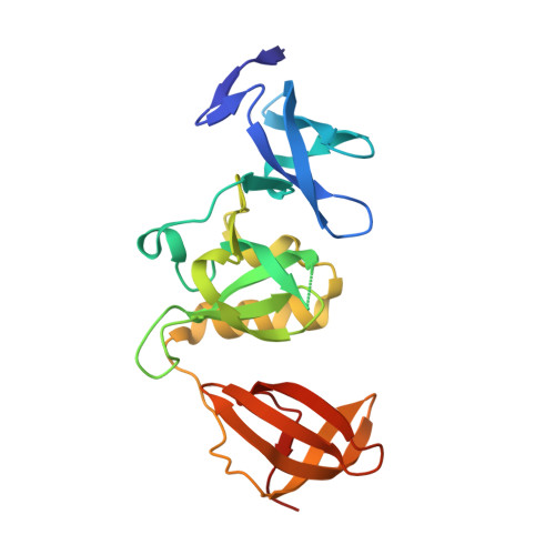
Crystal structure of SETDB1 Tudor domain with aryl triazole fragments


Experimental Data Snapshot

  • Method: X-RAY DIFFRACTION
  • Resolution: 1.80 Å
  • R-Value Free: 
    0.190 (Depositor), 0.193 (DCC) 
  • R-Value Work: 
    0.179 (Depositor) 
  • R-Value Observed: 
    0.179 (Depositor) 

wwPDB Validation 3D Report Full Report

Validation slider image for 6AU3

Ligand Structure Quality Assessment 


This is version 1.1 of the entry. See complete history

Macromolecule Content 

  • Total Structure Weight: 25.58 kDa 
  • Atom Count: 1,864 
  • Modeled Residue Count: 206 
  • Deposited Residue Count: 213 
  • Unique protein chains: 1

Macromolecules

Find similar proteins by:|  3D Structure
Entity ID: 1
MoleculeChains  Sequence LengthOrganismDetailsImage
Histone-lysine N-methyltransferase SETDB1213Homo sapiensMutation(s): 0 
Gene Names: SETDB1KIAA0067KMT1E
EC: 2.1.1.43 (PDB Primary Data), 2.1.1.366 (UniProt)
UniProt & NIH Common Fund Data Resources
Find proteins for Q15047 (Homo sapiens)
Explore Q15047 
Go to UniProtKB:  Q15047
PHAROS:  Q15047
GTEx:  ENSG00000143379 
Entity Groups
Sequence Clusters30% Identity50% Identity70% Identity90% Identity95% Identity100% Identity
UniProt GroupQ15047
Sequence Annotations
Expand
Reference Sequence

Small Molecules

Ligands 4 Unique
IDChains Name / Formula / InChI Key2D Diagram3D Interactions
BWM

Query on BWM


Download:Ideal Coordinates CCD File
B [auth A]N-{[2-(3,5-dimethyl-4H-1,2,4-triazol-4-yl)phenyl]methyl}acetamide
C13 H16 N4 O
FLTCDBPTYKZRMD-UHFFFAOYSA-N
SO4

Query on SO4


Download:Ideal Coordinates CCD File
C [auth A],
D [auth A],
E [auth A],
F [auth A]
SULFATE ION
O4 S
QAOWNCQODCNURD-UHFFFAOYSA-L
EDO

Query on EDO


Download:Ideal Coordinates CCD File
G [auth A]1,2-ETHANEDIOL
C2 H6 O2
LYCAIKOWRPUZTN-UHFFFAOYSA-N
UNX

Query on UNX


Download:Ideal Coordinates CCD File
H [auth A]
I [auth A]
J [auth A]
K [auth A]
L [auth A]
H [auth A],
I [auth A],
J [auth A],
K [auth A],
L [auth A],
M [auth A],
N [auth A]
UNKNOWN ATOM OR ION
X

Experimental Data & Validation

Experimental Data

  • Method: X-RAY DIFFRACTION
  • Resolution: 1.80 Å
  • R-Value Free:  0.190 (Depositor), 0.193 (DCC) 
  • R-Value Work:  0.179 (Depositor) 
  • R-Value Observed: 0.179 (Depositor) 
Space Group: P 21 21 21
Unit Cell:
Length ( Å )Angle ( ˚ )
a = 55.172α = 90
b = 63.528β = 90
c = 69.987γ = 90
Software Package:
Software NamePurpose
SCALEPACKdata scaling
REFMACrefinement
PDB_EXTRACTdata extraction
Cootmodel building
HKL-3000data reduction

Structure Validation

View Full Validation Report


Ligand Structure Quality Assessment 


Entry History 

Revision History  (Full details and data files)

  • Version 1.0: 2017-09-27
    Type: Initial release
  • Version 1.1: 2024-03-13
    Changes: Data collection, Database references