6ARK | pdb_00006ark

Crystal Structure of compound 10 covalently bound to K-Ras G12C


Experimental Data Snapshot

  • Method: X-RAY DIFFRACTION
  • Resolution: 1.75 Å
  • R-Value Free: 
    0.231 (Depositor), 0.234 (DCC) 
  • R-Value Work: 
    0.194 (Depositor), 0.198 (DCC) 
  • R-Value Observed: 
    0.196 (Depositor) 

Starting Model: experimental
View more details

wwPDB Validation 3D Report Full Report

Validation slider image for 6ARK

Ligand Structure Quality Assessment 


This is version 1.4 of the entry. See complete history

Literature

Novel K-Ras G12C Switch-II Covalent Binders Destabilize Ras and Accelerate Nucleotide Exchange.

Nnadi, C.I.Jenkins, M.L.Gentile, D.R.Bateman, L.A.Zaidman, D.Balius, T.E.Nomura, D.K.Burke, J.E.Shokat, K.M.London, N.

(2018) J Chem Inf Model 58: 464-471

  • DOI: https://doi.org/10.1021/acs.jcim.7b00399
  • Primary Citation Related Structures: 
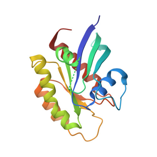
    6ARK

  • PubMed Abstract: 

    The success of targeted covalent inhibitors in the global pharmaceutical industry has led to a resurgence of covalent drug discovery. However, covalent inhibitor design for flexible binding sites remains a difficult task due to a lack of methodological development. Here, we compared covalent docking to empirical electrophile screening against the highly dynamic target K-Ras G12C . While the overall hit rate of both methods was comparable, we were able to rapidly progress a docking hit to a potent irreversible covalent binder that modifies the inactive, GDP-bound state of K-Ras G12C . Hydrogen-deuterium exchange mass spectrometry was used to probe the protein dynamics of compound binding to the switch-II pocket and subsequent destabilization of the nucleotide-binding region. SOS-mediated nucleotide exchange assays showed that, contrary to prior switch-II pocket inhibitors, these new compounds appear to accelerate nucleotide exchange. This study highlights the efficiency of covalent docking as a tool for the discovery of chemically novel hits against challenging targets.


  • Organizational Affiliation
    • Department of Cellular and Molecular Pharmacology, Howard Hughes Medical Institute, University of California, San Francisco , San Francisco, California 94158, United States.

Macromolecule Content 

  • Total Structure Weight: 20.4 kDa 
  • Atom Count: 1,444 
  • Modeled Residue Count: 166 
  • Deposited Residue Count: 170 
  • Unique protein chains: 1

Macromolecules

Find similar proteins by:|  3D Structure
Entity ID: 1
MoleculeChains  Sequence LengthOrganismDetailsImage
GTPase KRas170Homo sapiensMutation(s): 4 
Gene Names: KRASKRAS2RASK2
EC: 3.6.5.2
UniProt & NIH Common Fund Data Resources
Find proteins for P01116 (Homo sapiens)
Explore P01116 
Go to UniProtKB:  P01116
PHAROS:  P01116
GTEx:  ENSG00000133703 
Entity Groups
Sequence Clusters30% Identity50% Identity70% Identity90% Identity95% Identity100% Identity
UniProt GroupP01116
Sequence Annotations
Expand
Reference Sequence

Small Molecules

Ligands 4 Unique
IDChains Name / Formula / InChI Key2D Diagram3D Interactions
GDP

Query on GDP


Download:Ideal Coordinates CCD File
B [auth A]GUANOSINE-5'-DIPHOSPHATE
C10 H15 N5 O11 P2
QGWNDRXFNXRZMB-UUOKFMHZSA-N
BQD
(Subject of Investigation/LOI)

Query on BQD


Download:Ideal Coordinates CCD File
D [auth A](3R)-N-(6-bromonaphthalen-2-yl)-3-hydroxy-1-propanoyl-L-prolinamide
C18 H19 Br N2 O3
MFZHXIHGHGFXNJ-WBVHZDCISA-N
GOL

Query on GOL


Download:Ideal Coordinates CCD File
E [auth A],
F [auth A]
GLYCEROL
C3 H8 O3
PEDCQBHIVMGVHV-UHFFFAOYSA-N
MG

Query on MG


Download:Ideal Coordinates CCD File
C [auth A]MAGNESIUM ION
Mg
JLVVSXFLKOJNIY-UHFFFAOYSA-N

Experimental Data & Validation

Experimental Data

  • Method: X-RAY DIFFRACTION
  • Resolution: 1.75 Å
  • R-Value Free:  0.231 (Depositor), 0.234 (DCC) 
  • R-Value Work:  0.194 (Depositor), 0.198 (DCC) 
  • R-Value Observed: 0.196 (Depositor) 
Space Group: H 3 2
Unit Cell:
Length ( Å )Angle ( ˚ )
a = 94.28α = 90
b = 94.28β = 90
c = 119.55γ = 120
Software Package:
Software NamePurpose
PHENIXrefinement
Aimlessdata scaling
PDB_EXTRACTdata extraction
MOSFLMdata reduction
PHASERphasing

Structure Validation

View Full Validation Report


Ligand Structure Quality Assessment 


Entry History 

& Funding Information

Deposition Data


Funding OrganizationLocationGrant Number
National Institutes of Health/National Cancer Institute (NIH/NCI)United States5R01CA190408-03
Israel Science FoundationIsrael1097/16
National Institutes of Health/National Cancer Institute (NIH/NCI)United States1F30CA214015-01

Revision History  (Full details and data files)

  • Version 1.0: 2018-01-31
    Type: Initial release
  • Version 1.1: 2018-03-07
    Changes: Database references
  • Version 1.2: 2019-12-04
    Changes: Author supporting evidence
  • Version 1.3: 2023-10-04
    Changes: Data collection, Database references, Derived calculations, Refinement description
  • Version 1.4: 2024-10-16
    Changes: Structure summary