6ANI | pdb_00006ani

Coltuximab Fab in complex with anti-Kappa VHH domain


Experimental Data Snapshot

  • Method: X-RAY DIFFRACTION
  • Resolution: 2.40 Å
  • R-Value Free: 
    0.286 (Depositor), 0.288 (DCC) 
  • R-Value Work: 
    0.245 (Depositor), 0.247 (DCC) 
  • R-Value Observed: 
    0.247 (Depositor) 

wwPDB Validation 3D Report Full Report

Validation slider image for 6ANI

This is version 1.3 of the entry. See complete history

Literature

Structural Basis of Enhanced Crystallizability Induced by a Molecular Chaperone for Antibody Antigen-Binding Fragments.

Ereno-Orbea, J.Sicard, T.Cui, H.Carson, J.Hermans, P.Julien, J.P.

(2018) J Mol Biology 430: 322-336

  • DOI: https://doi.org/10.1016/j.jmb.2017.12.010
  • Primary Citation Related Structures: 
    6ANA, 6AND, 6ANI

  • PubMed Abstract: 

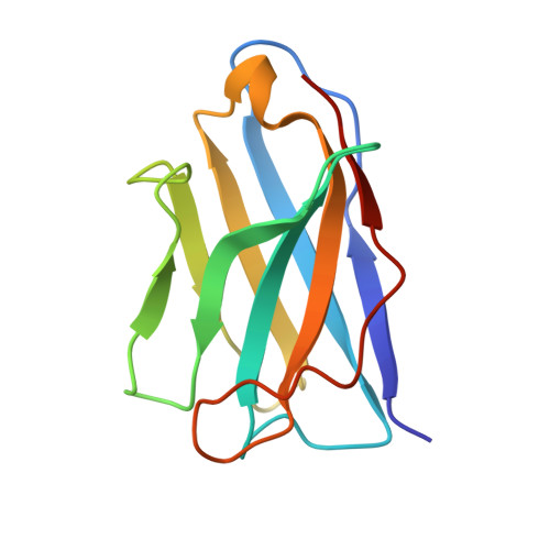
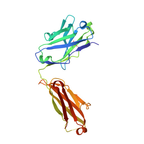
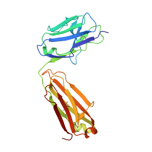
    Monoclonal antibodies constitute one of the largest groups of drugs to treat cancers and immune disorders, and are guiding the design of vaccines against infectious diseases. Fragments antigen-binding (Fabs) have been preferred over monoclonal antibodies for the structural characterization of antibody-antigen complexes due to their relatively low flexibility. Nonetheless, Fabs often remain challenging to crystallize because of the surface characteristics of complementary determining regions and the residual flexibility in the hinge region between the variable and constant domains. Here, we used a variable heavy-chain (V H H) domain specific for the human kappa light chain to assist in the structure determination of three therapeutic Fabs that were recalcitrant to crystallization on their own. We show that this ligand alters the surface properties of the antibody-ligand complex and lowers its aggregation temperature to favor crystallization. The V H H crystallization chaperone also restricts the flexible hinge of Fabs to a narrow range of angles, and so independently of the variable region. Our findings contribute a valuable approach to antibody structure determination and provide biophysical insight into the principles that govern the crystallization of macromolecules.


  • Organizational Affiliation
    • Program in Molecular Medicine, The Hospital for Sick Children Research Institute, Toronto, ON, Canada M5G 0A4.

Macromolecule Content 

  • Total Structure Weight: 114.58 kDa 
  • Atom Count: 7,823 
  • Modeled Residue Count: 1,088 
  • Deposited Residue Count: 1,110 
  • Unique protein chains: 3

Macromolecules

Find similar proteins by:|  3D Structure
Entity ID: 1
MoleculeChains  Sequence LengthOrganismDetailsImage
Anti-Kappa VHH domainA,
B [auth K]
121Lama glamaMutation(s): 0 
Find similar proteins by:|  3D Structure
Entity ID: 2
MoleculeChains  Sequence LengthOrganismDetailsImage
Coltuximab Fab heavy chainC [auth H],
E [auth I]
223Homo sapiensMutation(s): 0 
Find similar proteins by:|  3D Structure
Entity ID: 3
MoleculeChains  Sequence LengthOrganismDetailsImage
Coltuximab Fab light chainD [auth L],
F [auth M]
211Homo sapiensMutation(s): 0 

Experimental Data & Validation

Experimental Data

  • Method: X-RAY DIFFRACTION
  • Resolution: 2.40 Å
  • R-Value Free:  0.286 (Depositor), 0.288 (DCC) 
  • R-Value Work:  0.245 (Depositor), 0.247 (DCC) 
  • R-Value Observed: 0.247 (Depositor) 
Space Group: C 1 2 1
Unit Cell:
Length ( Å )Angle ( ˚ )
a = 182.39α = 90
b = 48.301β = 104.07
c = 128.547γ = 90
Software Package:
Software NamePurpose
PHENIXrefinement
XSCALEdata scaling
PDB_EXTRACTdata extraction
XDSdata reduction
PHASERphasing

Structure Validation

View Full Validation Report


Entry History 

Deposition Data

Revision History  (Full details and data files)

  • Version 1.0: 2018-01-31
    Type: Initial release
  • Version 1.1: 2018-02-14
    Changes: Database references
  • Version 1.2: 2019-10-16
    Changes: Advisory, Data collection, Structure summary
  • Version 1.3: 2024-11-20
    Changes: Data collection, Database references, Structure summary