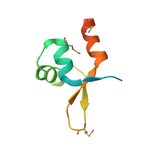
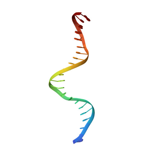
6AMK | pdb_00006amk

Structure of Streptomyces venezuelae BldC-whiI opt complex


Experimental Data Snapshot

  • Method: X-RAY DIFFRACTION
  • Resolution: 3.29 Å
  • R-Value Free: 
    0.263 (Depositor), 0.282 (DCC) 
  • R-Value Work: 
    0.223 (Depositor), 0.240 (DCC) 
  • R-Value Observed: 
    0.227 (Depositor) 

wwPDB Validation 3D Report Full Report

Validation slider image for 6AMK

This is version 1.3 of the entry. See complete history. 

Literature

The MerR-like protein BldC binds DNA direct repeats as cooperative multimers to regulate Streptomyces development.

Schumacher, M.A., den Hengst, C.D., Bush, M.J., Le, T.B.K., Tran, N.T., Chandra, G., Zeng, W., Travis, B., Brennan, R.G., Buttner, M.J.

(2018) Nat Commun 9: 1139-1139

  • DOI: https://doi.org/10.1038/s41467-018-03576-3
  • Primary Citation Related Structures: 
    6AMA, 6AMK

  • PubMed Abstract: 

    Streptomycetes are notable for their complex life cycle and production of most clinically important antibiotics. A key factor that controls entry into development and the onset of antibiotic production is the 68-residue protein, BldC. BldC is a putative DNA-binding protein related to MerR regulators, but lacks coiled-coil dimerization and effector-binding domains characteristic of classical MerR proteins. Hence, the molecular function of the protein has been unclear. Here we show that BldC is indeed a DNA-binding protein and controls a regulon that includes other key developmental regulators. Intriguingly, BldC DNA-binding sites vary significantly in length. Our BldC-DNA structures explain this DNA-binding capability by revealing that BldC utilizes a DNA-binding mode distinct from MerR and other known regulators, involving asymmetric head-to-tail oligomerization on DNA direct repeats that results in dramatic DNA distortion. Notably, BldC-like proteins radiate throughout eubacteria, establishing BldC as the founding member of a new structural family of regulators.


  • Organizational Affiliation: 
    • Department of Biochemistry, Duke University School of Medicine, Durham, NC, 27710, USA. maria.schumacher@duke.edu.

Macromolecule Content 

  • Total Structure Weight: 29.84 kDa 
  • Atom Count: 1,698 
  • Modeled Residue Count: 147 
  • Deposited Residue Count: 188 
  • Unique protein chains: 1
  • Unique nucleic acid chains: 2

Macromolecules


Find similar proteins by:|  3D Structure
Entity ID: 1
MoleculeChains  Sequence LengthOrganismDetailsImage
Putative DNA-binding protein
A, B
72Streptomyces venezuelaeMutation(s): 2 
Gene Names: AQF52_4259
UniProt
Find proteins for F2REK9 (Streptomyces venezuelae (strain ATCC 10712 / CBS 650.69 / DSM 40230 / JCM 4526 / NBRC 13096 / PD 04745))
Explore F2REK9 
Go to UniProtKB:  F2REK9
Entity Groups
Sequence Clusters30% Identity50% Identity70% Identity90% Identity95% Identity100% Identity
UniProt GroupF2REK9
Sequence Annotations
Expand
Reference Sequence
Find similar nucleic acids by:  Sequence
Entity ID: 2
MoleculeChains LengthOrganismImage
DNA (5'-D(*TP*TP*CP*AP*AP*TP*TP*CP*GP*GP*GP*TP*AP*AP*TP*TP*CP*GP*GP*GP*CP*A)-3')C [auth R]22synthetic construct
Sequence Annotations
Expand
Reference Sequence
Find similar nucleic acids by:  Sequence
Entity ID: 3
MoleculeChains LengthOrganismImage
DNA (5'-D(*AP*AP*TP*GP*TP*CP*CP*GP*AP*AP*TP*TP*AP*CP*CP*CP*GP*AP*AP*TP*TP*G)-3')D [auth Z]22synthetic construct
Sequence Annotations
Expand
Reference Sequence

Small Molecules

Modified Residues  1 Unique
IDChains TypeFormula2D DiagramParent
MSE
Query on MSE
A, B
L-PEPTIDE LINKINGC5 H11 N O2 SeMET

Experimental Data & Validation

Experimental Data

  • Method: X-RAY DIFFRACTION
  • Resolution: 3.29 Å
  • R-Value Free:  0.263 (Depositor), 0.282 (DCC) 
  • R-Value Work:  0.223 (Depositor), 0.240 (DCC) 
  • R-Value Observed: 0.227 (Depositor) 
Space Group: P 61 2 2
Unit Cell:
Length ( Å )Angle ( ˚ )
a = 114.2α = 90
b = 114.2β = 90
c = 159γ = 120
Software Package:
Software NamePurpose
MOSFLMdata collection
SCALAdata scaling
PHASERphasing
PHENIXrefinement
PDB_EXTRACTdata extraction

Structure Validation

View Full Validation Report


Entry History 

Deposition Data

Revision History  (Full details and data files)

  • Version 1.0: 2018-03-28
    Type: Initial release
  • Version 1.1: 2018-04-04
    Changes: Data collection, Database references
  • Version 1.2: 2018-11-07
    Changes: Data collection, Source and taxonomy
  • Version 1.3: 2024-10-16
    Changes: Data collection, Database references, Structure summary