6AIY

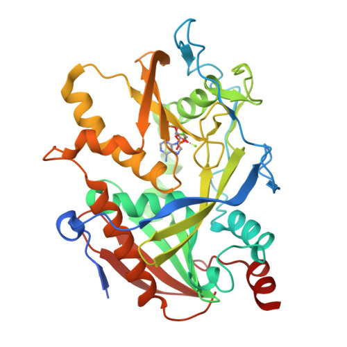
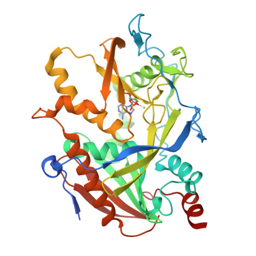
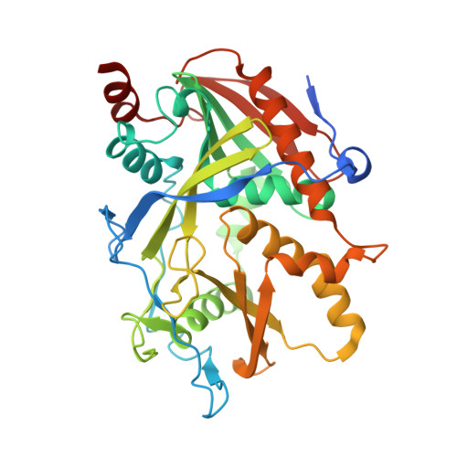
Crystal structure of DXO (E234A mutant) in complex with adenosine 3', 5' bisphosphate and two magnesium ions


Experimental Data Snapshot

  • Method: X-RAY DIFFRACTION
  • Resolution: 1.90 Å
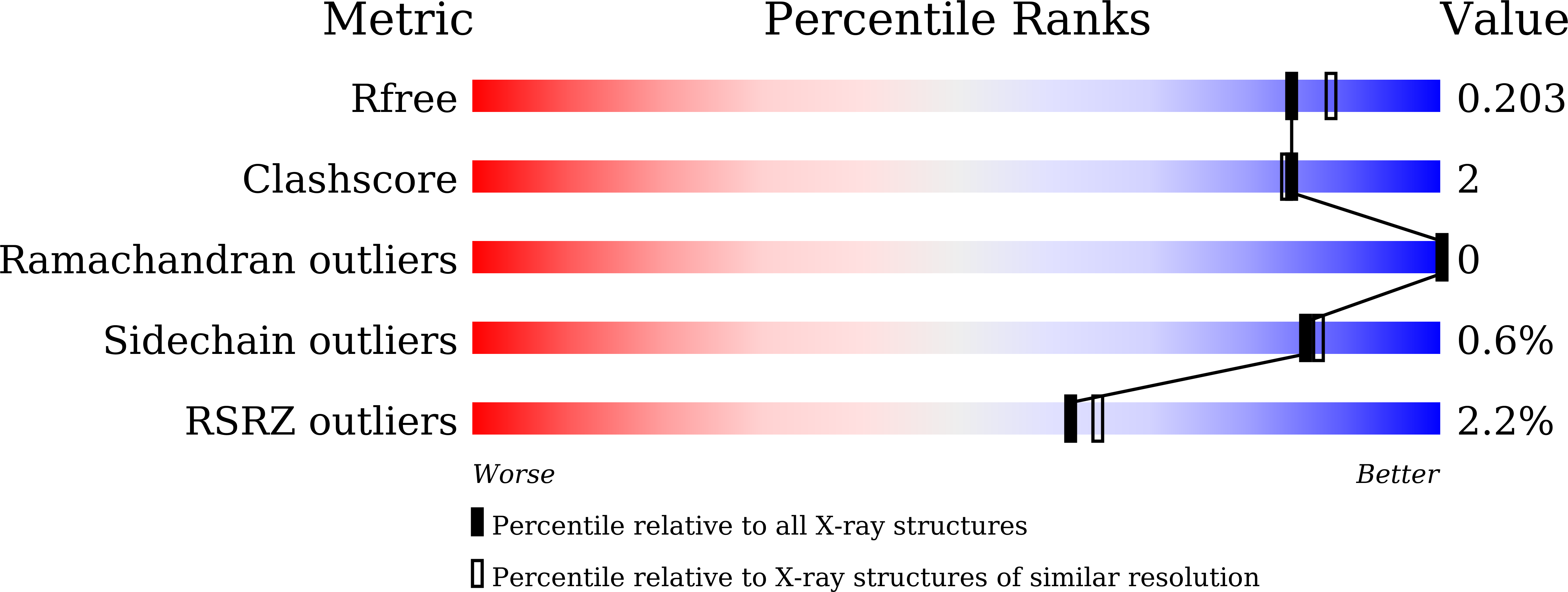
  • R-Value Free: 0.203 
  • R-Value Work: 0.170 
  • R-Value Observed: 0.172 

wwPDB Validation   3D Report Full Report


Ligand Structure Quality Assessment 


This is version 1.2 of the entry. See complete history


Literature

Molecular mechanism for the inhibition of DXO by adenosine 3',5'-bisphosphate.

Yun, J.S.Yoon, J.H.Choi, Y.J.Son, Y.J.Kim, S.Tong, L.Chang, J.H.

(2018) Biochem Biophys Res Commun 504: 89-95

  • DOI: https://doi.org/10.1016/j.bbrc.2018.08.135
  • Primary Citation of Related Structures:  
    6AIX, 6AIY

  • PubMed Abstract: 

    The decapping exoribonuclease DXO functions in pre-mRNA capping quality control, and shows multiple biochemical activities such as decapping, deNADding, pyrophosphohydrolase, and 5'-3' exoribonuclease activities. Previous studies revealed the molecular mechanisms of DXO based on the structures in complexes with a product, substrate mimic, cap analogue, and 3'-NADP + . Despite several reports on the substrate-specific reaction mechanism, the inhibitory mechanism of DXO remains elusive. Here, we demonstrate that adenosine 3', 5'-bisphosphate (pAp), a known inhibitor of the 5'-3' exoribonuclease Xrn1, inhibits the nuclease activity of DXO based on the results of structural and biochemical experiments. We determined the crystal structure of the DXO-pAp-Mg 2+ complex at 1.8 Å resolution. In comparison with the DXO-RNA product complex, the position of pAp is well superimposed with the first nucleotide of the product RNA in the vicinity of two magnesium ions. Furthermore, biochemical assays showed that the inhibition by pAp is comparable between Xrn1 and DXO. Collectively, these structural and biochemical studies reveal that pAp inhibits the activities of DXO by occupying the active site to act as a competitive inhibitor.


  • Organizational Affiliation

    Department of Biology Education, Kyungpook National University, Daegu 41566, South Korea.


Macromolecules
Find similar proteins by:  (by identity cutoff)  |  3D Structure
Entity ID: 1
MoleculeChains Sequence LengthOrganismDetailsImage
Decapping and exoribonuclease protein378Mus musculusMutation(s): 1 
Gene Names: DxoDom3zNg6
EC: 3.1.13 (PDB Primary Data), 3.6.1 (PDB Primary Data)
UniProt
Find proteins for O70348 (Mus musculus)
Explore O70348 
Go to UniProtKB:  O70348
Entity Groups  
Sequence Clusters30% Identity50% Identity70% Identity90% Identity95% Identity100% Identity
UniProt GroupO70348
Sequence Annotations
Expand
  • Reference Sequence
Experimental Data & Validation

Experimental Data

  • Method: X-RAY DIFFRACTION
  • Resolution: 1.90 Å
  • R-Value Free: 0.203 
  • R-Value Work: 0.170 
  • R-Value Observed: 0.172 
  • Space Group: P 1 21 1
Unit Cell:
Length ( Å )Angle ( ˚ )
a = 46.499α = 90
b = 88.081β = 114.42
c = 50.04γ = 90
Software Package:
Software NamePurpose
PHENIXrefinement
HKL-2000data reduction
HKL-2000data scaling
CNSphasing

Structure Validation

View Full Validation Report


Ligand Structure Quality Assessment 


Entry History & Funding Information

Deposition Data


Funding OrganizationLocationGrant Number
National Research Foundation (Korea)Korea, Republic Of2016R1C1B2009691
National Institutes of Health/National Institute of General Medical Sciences (NIH/NIGMS)United StatesGM118093

Revision History  (Full details and data files)

  • Version 1.0: 2018-10-17
    Type: Initial release
  • Version 1.1: 2022-03-23
    Changes: Author supporting evidence, Database references, Derived calculations
  • Version 1.2: 2024-05-29
    Changes: Data collection