6AIG | pdb_00006aig

Crystal structure of PCNA1 from Aeropyrum pernix


Experimental Data Snapshot

  • Method: X-RAY DIFFRACTION
  • Resolution: 2.00 Å
  • R-Value Free: 
    0.226 (Depositor), 0.226 (DCC) 
  • R-Value Work: 
    0.185 (Depositor), 0.193 (DCC) 
  • R-Value Observed: 
    0.187 (Depositor) 

Starting Model: experimental
View more details

wwPDB Validation 3D Report Full Report

Validation slider image for 6AIG

This is version 1.3 of the entry. See complete history

Literature

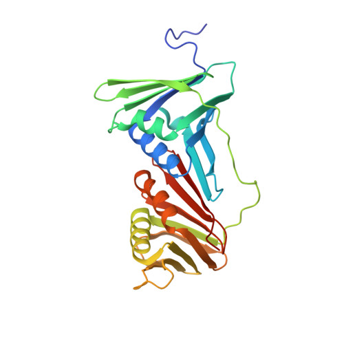
X-ray crystal structure of proliferating cell nuclear antigen 1 from Aeropyrum pernix.

Yamauchi, T.Kikuchi, M.Iizuka, Y.Tsunoda, M.

(2024) Acta Crystallogr F Struct Biol Commun 80: 294-301

  • DOI: https://doi.org/10.1107/S2053230X24009518
  • Primary Citation Related Structures: 
    6AIG

  • PubMed Abstract: 

    Proliferating cell nuclear antigen (PCNA) plays a critical role in DNA replication by enhancing the activity of various proteins involved in replication. In this study, the crystal structure of ApePCNA1, one of three PCNAs from the thermophilic archaeon Aeropyrum pernix, was elucidated. ApePCNA1 was cloned and expressed in Escherichia coli and the protein was purified and crystallized. The resulting crystal structure determined at 2.00 Å resolution revealed that ApePCNA1 does not form a trimeric ring, unlike PCNAs from other domains of life. It has unique structural features, including a long interdomain-connecting loop and a PIP-box-like sequence at the N-terminus, indicating potential interactions with other proteins. These findings provide insights into the functional mechanisms of PCNAs in archaea and their evolutionary conservation across different domains of life. A modified medium and protocol were used to express recombinant protein containing the lac operon. The expression of the target protein increased and the total incubation time decreased when using this system compared with those of previous expression protocols.


  • Organizational Affiliation
    • Graduate School of Life Science and Technology, Iryo Sosei University, Iwaki, Fukushima, Japan.

Macromolecule Content 

  • Total Structure Weight: 29.48 kDa 
  • Atom Count: 2,049 
  • Modeled Residue Count: 258 
  • Deposited Residue Count: 263 
  • Unique protein chains: 1

Macromolecules

Find similar proteins by:|  3D Structure
Entity ID: 1
MoleculeChains  Sequence LengthOrganismDetailsImage
DNA polymerase sliding clamp 1263Aeropyrum pernix K1Mutation(s): 0 
Gene Names: APE_0162
UniProt
Find proteins for Q9YFT8 (Aeropyrum pernix (strain ATCC 700893 / DSM 11879 / JCM 9820 / NBRC 100138 / K1))
Explore Q9YFT8 
Go to UniProtKB:  Q9YFT8
Entity Groups
Sequence Clusters30% Identity50% Identity70% Identity90% Identity95% Identity100% Identity
UniProt GroupQ9YFT8
Sequence Annotations
Expand
Reference Sequence

Experimental Data & Validation

Experimental Data

  • Method: X-RAY DIFFRACTION
  • Resolution: 2.00 Å
  • R-Value Free:  0.226 (Depositor), 0.226 (DCC) 
  • R-Value Work:  0.185 (Depositor), 0.193 (DCC) 
  • R-Value Observed: 0.187 (Depositor) 
Space Group: P 43 21 2
Unit Cell:
Length ( Å )Angle ( ˚ )
a = 69.028α = 90
b = 69.028β = 90
c = 120.857γ = 90
Software Package:
Software NamePurpose
REFMACrefinement
XDSdata reduction
XDSdata scaling
MOLREPphasing
ARP/wARPmodel building

Structure Validation

View Full Validation Report


Entry History 

Deposition Data

Revision History  (Full details and data files)

  • Version 1.0: 2018-09-26
    Type: Initial release
  • Version 1.1: 2023-06-28
    Changes: Database references, Refinement description
  • Version 1.2: 2023-11-29
    Changes: Data collection, Refinement description
  • Version 1.3: 2024-11-13
    Changes: Database references, Structure summary