6AH7 | pdb_00006ah7

D45W/H226G mutant of marine bacterial prolidase


Experimental Data Snapshot

  • Method: X-RAY DIFFRACTION
  • Resolution: 2.38 Å
  • R-Value Free: 
    0.269 (Depositor), 0.271 (DCC) 
  • R-Value Work: 
    0.233 (Depositor), 0.237 (DCC) 

Starting Model: experimental
View more details

wwPDB Validation 3D Report Full Report

Validation slider image for 6AH7

This is version 1.4 of the entry. See complete history

Literature

Repurposing a bacterial prolidase for organophosphorus hydrolysis: Reshaped catalytic cavity switches substrate selectivity.

Yang, J.Xiao, Y.Z.Li, R.Liu, Y.Long, L.J.

(2020) Biotechnol Bioeng 117: 2694-2702

  • DOI: https://doi.org/10.1002/bit.27455
  • Primary Citation Related Structures: 
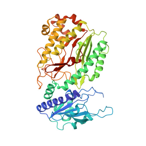
    6AH7, 6AH8

  • PubMed Abstract: 

    Enzyme promiscuity is critical to the acquisition of evolutionary plasticity in cells and can be recruited for high-value chemical synthesis or xenobiotic degradation. The molecular determinants of substrate ambiguity are essential to this activity; however, these details remain unknown. Here, we performed the directed evolution of a prolidase to enhance its initially weak paraoxonase activity. The in vitro evolution led to an unexpected 1,000,000-fold switch in substrate selectivity, with a 30-fold increase in paraoxon hydrolysis and 40,000-fold decrease in peptide hydrolysis. Structural and in silico analyses revealed enlarged catalytic cavities and substrate repositioning as responsible for rapid catalytic transitions between distinct chemical reactions.


  • Organizational Affiliation
    • CAS Key Laboratory of Tropical Marine Bio-resources and Ecology, Guangdong Key Laboratory of Marine Materia Medica, South China Sea Institute of Oceanology, Chinese Academy of Sciences, Guangzhou, China.

Macromolecule Content 

  • Total Structure Weight: 207.08 kDa 
  • Atom Count: 14,924 
  • Modeled Residue Count: 1,756 
  • Deposited Residue Count: 1,792 
  • Unique protein chains: 1

Macromolecules

Find similar proteins by:|  3D Structure
Entity ID: 1
MoleculeChains  Sequence LengthOrganismDetailsImage
Xaa-Pro dipeptidase
A, B, C, D
448Pseudoalteromonas lipolyticaMutation(s): 2 
Gene Names: pepQSAMN04487854_12236
EC: 3.4.13.9

Small Molecules

Ligands 3 Unique
IDChains Name / Formula / InChI Key2D Diagram3D Interactions
SO4

Query on SO4


Download:Ideal Coordinates CCD File
G [auth A]SULFATE ION
O4 S
QAOWNCQODCNURD-UHFFFAOYSA-L
MN

Query on MN


Download:Ideal Coordinates CCD File
E [auth A]
F [auth A]
I [auth B]
J [auth B]
K [auth C]
E [auth A],
F [auth A],
I [auth B],
J [auth B],
K [auth C],
L [auth C],
M [auth D],
N [auth D]
MANGANESE (II) ION
Mn
WAEMQWOKJMHJLA-UHFFFAOYSA-N
NA

Query on NA


Download:Ideal Coordinates CCD File
H [auth A]SODIUM ION
Na
FKNQFGJONOIPTF-UHFFFAOYSA-N

Experimental Data & Validation

Experimental Data

  • Method: X-RAY DIFFRACTION
  • Resolution: 2.38 Å
  • R-Value Free:  0.269 (Depositor), 0.271 (DCC) 
  • R-Value Work:  0.233 (Depositor), 0.237 (DCC) 
Space Group: H 3 2
Unit Cell:
Length ( Å )Angle ( ˚ )
a = 183.632α = 90
b = 183.632β = 90
c = 371.306γ = 120
Software Package:
Software NamePurpose
PHENIXrefinement
XDSdata reduction
XDSdata scaling
PHASERphasing

Structure Validation

View Full Validation Report


Entry History 

Deposition Data

Revision History  (Full details and data files)

  • Version 1.0: 2019-09-18
    Type: Initial release
  • Version 1.1: 2019-09-25
    Changes: Data collection, Derived calculations
  • Version 1.2: 2019-11-20
    Changes: Derived calculations
  • Version 1.3: 2020-09-23
    Changes: Database references, Derived calculations
  • Version 1.4: 2023-11-22
    Changes: Data collection, Database references, Refinement description