6AG5 | pdb_00006ag5

Crystal structure of Ard1 N-terminal acetyltransferase E88H/H127E mutant from Sulfolobus solfataricus


Experimental Data Snapshot

  • Method: X-RAY DIFFRACTION
  • Resolution: 2.32 Å
  • R-Value Free: 
    0.249 (Depositor), 0.251 (DCC) 
  • R-Value Work: 
    0.160 (Depositor), 0.169 (DCC) 
  • R-Value Observed: 
    0.169 (Depositor) 

Starting Model: experimental
View more details

wwPDB Validation 3D Report Full Report

Validation slider image for 6AG5

Ligand Structure Quality Assessment 


This is version 1.2 of the entry. See complete history

Literature

Adaptation of thermophilic acetyltransferase to a water-mediated catalytic mechanism.

Chang, Y.Y.Hagawa, S.Hsu, C.H.

(2020) Chem Commun (Camb) 56: 10537-10540

  • DOI: https://doi.org/10.1039/d0cc04305b
  • Primary Citation Related Structures: 
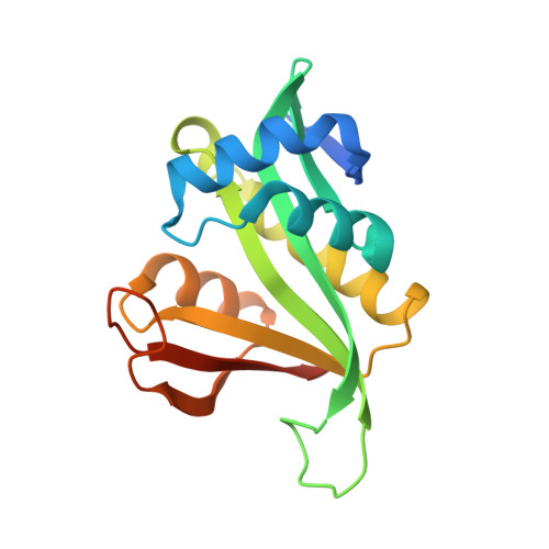
    6AG4, 6AG5

  • PubMed Abstract: 

    The common mechanism of N-acetyltransferases (NATs) is a water-mediated catalysis, which is not conducive to thermophilic acetyltransferases. The crystal structure of SsArd1 shows an ordered catalytic water molecule in a trap formed by the residues H88 and E127. Structure-guided mutagenesis, kinetic studies and MD simulation indicated that the turnover rates of H88A, E127A and H88A/E127A mutants were low, but that of the H88E/E127H mutant could be restored to the level of the wild type.


  • Organizational Affiliation
    • Department of Agricultural Chemistry, National Taiwan University, Taipei 10617, Taiwan. andyhsu@ntu.edu.tw.

Macromolecule Content 

  • Total Structure Weight: 21.36 kDa 
  • Atom Count: 1,443 
  • Modeled Residue Count: 157 
  • Deposited Residue Count: 175 
  • Unique protein chains: 1

Macromolecules

Find similar proteins by:|  3D Structure
Entity ID: 1
MoleculeChains  Sequence LengthOrganismDetailsImage
N-alpha-acetyltransferase175Saccharolobus solfataricus P2Mutation(s): 2 
Gene Names: AsArd1
EC: 2.3.1.255 (PDB Primary Data), 2.3.1.258 (PDB Primary Data)
UniProt
Find proteins for Q980R9 (Saccharolobus solfataricus (strain ATCC 35092 / DSM 1617 / JCM 11322 / P2))
Explore Q980R9 
Go to UniProtKB:  Q980R9
Entity Groups
Sequence Clusters30% Identity50% Identity70% Identity90% Identity95% Identity100% Identity
UniProt GroupQ980R9
Sequence Annotations
Expand
Reference Sequence

Experimental Data & Validation

Experimental Data

  • Method: X-RAY DIFFRACTION
  • Resolution: 2.32 Å
  • R-Value Free:  0.249 (Depositor), 0.251 (DCC) 
  • R-Value Work:  0.160 (Depositor), 0.169 (DCC) 
  • R-Value Observed: 0.169 (Depositor) 
Space Group: P 21 21 21
Unit Cell:
Length ( Å )Angle ( ˚ )
a = 41.217α = 90
b = 52.926β = 90
c = 73.576γ = 90
Software Package:
Software NamePurpose
PHENIXrefinement
HKL-2000data reduction
HKL-2000data scaling
PHASERphasing

Structure Validation

View Full Validation Report


Ligand Structure Quality Assessment 


Entry History 

& Funding Information

Deposition Data


Funding OrganizationLocationGrant Number
Ministry of Science and Technology (Taiwan)Taiwan103-2113-M-002 -009 -MY2

Revision History  (Full details and data files)

  • Version 1.0: 2019-08-14
    Type: Initial release
  • Version 1.1: 2020-09-23
    Changes: Database references
  • Version 1.2: 2023-11-22
    Changes: Data collection, Database references, Refinement description