6A9W | pdb_00006a9w

Structure of the bifunctional DNA primase-polymerase from phage NrS-1


Experimental Data Snapshot

  • Method: X-RAY DIFFRACTION
  • Resolution: 1.80 Å
  • R-Value Free: 
    0.218 (Depositor), 0.220 (DCC) 
  • R-Value Work: 
    0.178 (Depositor), 0.183 (DCC) 
  • R-Value Observed: 
    0.180 (Depositor) 

wwPDB Validation 3D Report Full Report

Validation slider image for 6A9W

This is version 1.1 of the entry. See complete history

Literature

Crystal structure and biochemical studies of the bifunctional DNA primase-polymerase from phage NrS-1.

Guo, H.J.Li, M.J.Wang, T.L.Wu, H.Zhou, H.Xu, C.Y.Liu, X.P.Yu, F.He, J.H.

(2019) Biochem Biophys Res Commun 510: 573-579

  • DOI: https://doi.org/10.1016/j.bbrc.2019.01.144
  • Primary Citation Related Structures: 
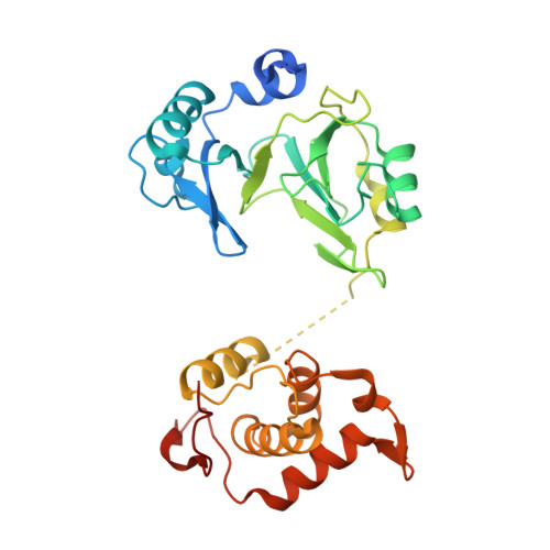
    6A9W

  • PubMed Abstract: 

    A novel DNA polymerase found in the deep-sea vent phage NrS-1, was confirmed to have both DNA polymerase and primase activities. In this polymerase, the N-terminal residues 1-300 (referred to as N300) are the core region required for polymerizing DNA and catalyzing de novo DNA synthesis. Here, the crystal structure of N300 was solved at a resolution of 1.80 Å. The overall structure consists of a prim/pol domain and a helix bundle domain, which are separated by a 14-residue-long flexible tether (residues 177-190). Both the prim/pol domain of N300 and other primase-polymerases (prim-pol) encompass an analogous fold with conserved catalytic residues. Mutagenesis and enzymatic activity assays show that the acidic active-site residue E139 is required for both polymerase and primase activities. Functional assays confirm the essentiality of the helix bundle domain for primase activity. Furthermore, we identified a mutant (N300-Y261A) of the helix bundle domain, which probably plays an indispensable role in the primer initiation and recognition of template DNA.


  • Organizational Affiliation
    • Shanghai Institute of Applied Physics, Chinese Academy of Sciences, Shanghai, 201800, China; University of Chinese Academy of Sciences, Beijing, 100049, China.

Macromolecule Content 

  • Total Structure Weight: 36.31 kDa 
  • Atom Count: 2,436 
  • Modeled Residue Count: 281 
  • Deposited Residue Count: 320 
  • Unique protein chains: 1

Macromolecules

Find similar proteins by:|  3D Structure
Entity ID: 1
MoleculeChains  Sequence LengthOrganismDetailsImage
Primase320Nitratiruptor phage NrS-1Mutation(s): 0 
EC: 2.7.7.7 (UniProt), 3.6.4.12 (UniProt), 2.7.7 (UniProt)
UniProt
Find proteins for M5AAG8 (Nitratiruptor phage NrS-1)
Explore M5AAG8 
Go to UniProtKB:  M5AAG8
Entity Groups
Sequence Clusters30% Identity50% Identity70% Identity90% Identity95% Identity100% Identity
UniProt GroupM5AAG8
Sequence Annotations
Expand
Reference Sequence

Experimental Data & Validation

Experimental Data

  • Method: X-RAY DIFFRACTION
  • Resolution: 1.80 Å
  • R-Value Free:  0.218 (Depositor), 0.220 (DCC) 
  • R-Value Work:  0.178 (Depositor), 0.183 (DCC) 
  • R-Value Observed: 0.180 (Depositor) 
Space Group: C 1 2 1
Unit Cell:
Length ( Å )Angle ( ˚ )
a = 119.874α = 90
b = 57.57β = 94.03
c = 47.631γ = 90
Software Package:
Software NamePurpose
PHENIXrefinement
XDSdata reduction
Aimlessdata scaling
PHASERphasing
PDB_EXTRACTdata extraction

Structure Validation

View Full Validation Report


Entry History 

& Funding Information

Deposition Data


Funding OrganizationLocationGrant Number
National Natural Science Foundation of ChinaChina31570740

Revision History  (Full details and data files)

  • Version 1.0: 2019-03-13
    Type: Initial release
  • Version 1.1: 2024-03-27
    Changes: Data collection, Database references