6A94

Crystal structure of 5-HT2AR in complex with zotepine


Experimental Data Snapshot

  • Method: X-RAY DIFFRACTION
  • Resolution: 2.90 Å
  • R-Value Free: 
    0.269 (Depositor), 0.270 (DCC) 
  • R-Value Work: 
    0.236 (Depositor), 0.240 (DCC) 
  • R-Value Observed: 
    0.238 (Depositor) 

Starting Model: experimental
View more details

wwPDB Validation   3D Report Full Report


Ligand Structure Quality Assessment 

Created with Raphaël 2.3.0Worse 01 BetterLigand structure goodness of fit to experimental dataBest fitted CLRClick on this verticalbar to view detailsBest fitted A6LClick on this verticalbar to view detailsBest fitted ZOTClick on this verticalbar to view detailsBest fitted PLMClick on this verticalbar to view details

This is version 1.3 of the entry. See complete history


Literature

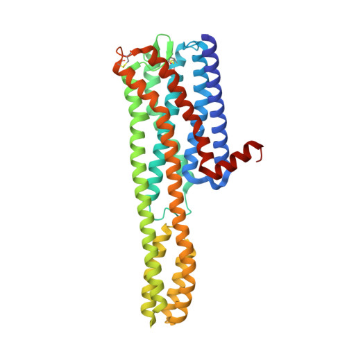
Structures of the 5-HT2Areceptor in complex with the antipsychotics risperidone and zotepine.

Kimura, K.T.Asada, H.Inoue, A.Kadji, F.M.N.Im, D.Mori, C.Arakawa, T.Hirata, K.Nomura, Y.Nomura, N.Aoki, J.Iwata, S.Shimamura, T.

(2019) Nat Struct Mol Biol 26: 121-128

  • DOI: https://doi.org/10.1038/s41594-018-0180-z
  • Primary Citation of Related Structures:  
    6A93, 6A94

  • PubMed Abstract: 

    Many drugs target the serotonin 2A receptor (5-HT 2A R), including second-generation antipsychotics that also target the dopamine D 2 receptor (D 2 R). These drugs often produce severe side effects due to non-selective binding to other aminergic receptors. Here, we report the structures of human 5-HT 2A R in complex with the second-generation antipsychotics risperidone and zotepine. These antipsychotics effectively stabilize the inactive conformation by forming direct contacts with the residues at the bottom of the ligand-binding pocket, the movements of which are important for receptor activation. 5-HT 2A R is structurally similar to 5-HT 2C R but possesses a unique side-extended cavity near the orthosteric binding site. A docking study and mutagenic studies suggest that a highly 5-HT 2A R-selective antagonist binds the side-extended cavity. The conformation of the ligand-binding pocket in 5-HT 2A R significantly differs around extracellular loops 1 and 2 from that in D 2 R. These findings are beneficial for the rational design of safer antipsychotics and 5-HT 2A R-selective drugs.


  • Organizational Affiliation

    Department of Cell Biology, Graduate School of Medicine, Kyoto University, Kyoto, Kyoto, Japan.


Macromolecules
Find similar proteins by:  (by identity cutoff)  |  3D Structure
Entity ID: 1
MoleculeChains Sequence LengthOrganismDetailsImage
5-hydroxytryptamine receptor 2A,Soluble cytochrome b562
A, B
376Homo sapiensEscherichia coliMutation(s): 6 
Gene Names: HTR2AHTR2cybC
Membrane Entity: Yes 
UniProt & NIH Common Fund Data Resources
Find proteins for P28223 (Homo sapiens)
Explore P28223 
Go to UniProtKB:  P28223
PHAROS:  P28223
GTEx:  ENSG00000102468 
Find proteins for P0ABE7 (Escherichia coli)
Explore P0ABE7 
Go to UniProtKB:  P0ABE7
Entity Groups  
Sequence Clusters30% Identity50% Identity70% Identity90% Identity95% Identity100% Identity
UniProt GroupsP28223P0ABE7
Sequence Annotations
Expand
  • Reference Sequence
Small Molecules
Ligands 6 Unique
IDChains Name / Formula / InChI Key2D Diagram3D Interactions
CLR
Query on CLR

Download Ideal Coordinates CCD File 
D [auth A],
I [auth B]
CHOLESTEROL
C27 H46 O
HVYWMOMLDIMFJA-DPAQBDIFSA-N
A6L
Query on A6L

Download Ideal Coordinates CCD File 
J [auth B]2,3-dihydroxypropyl (9Z)-octadec-9-enoate
C21 H40 O4
RZRNAYUHWVFMIP-KTKRTIGZSA-N
ZOT
Query on ZOT

Download Ideal Coordinates CCD File 
C [auth A],
G [auth B]
2-(3-chloranylbenzo[b][1]benzothiepin-5-yl)oxy-N,N-dimethyl-ethanamine
C18 H18 Cl N O S
HDOZVRUNCMBHFH-UHFFFAOYSA-N
PLM
Query on PLM

Download Ideal Coordinates CCD File 
H [auth B]PALMITIC ACID
C16 H32 O2
IPCSVZSSVZVIGE-UHFFFAOYSA-N
1PE
Query on 1PE

Download Ideal Coordinates CCD File 
E [auth A]PENTAETHYLENE GLYCOL
C10 H22 O6
JLFNLZLINWHATN-UHFFFAOYSA-N
ZN
Query on ZN

Download Ideal Coordinates CCD File 
F [auth A]ZINC ION
Zn
PTFCDOFLOPIGGS-UHFFFAOYSA-N
Experimental Data & Validation

Experimental Data

  • Method: X-RAY DIFFRACTION
  • Resolution: 2.90 Å
  • R-Value Free:  0.269 (Depositor), 0.270 (DCC) 
  • R-Value Work:  0.236 (Depositor), 0.240 (DCC) 
  • R-Value Observed: 0.238 (Depositor) 
Space Group: C 1 2 1
Unit Cell:
Length ( Å )Angle ( ˚ )
a = 279.18α = 90
b = 42.22β = 92.16
c = 91.71γ = 90
Software Package:
Software NamePurpose
PHENIXrefinement
XDSdata reduction
XSCALEdata scaling
PHASERphasing

Structure Validation

View Full Validation Report


Ligand Structure Quality Assessment 

Created with Raphaël 2.3.0Worse 01 BetterLigand structure goodness of fit to experimental dataBest fitted CLRClick on this verticalbar to view detailsBest fitted A6LClick on this verticalbar to view detailsBest fitted ZOTClick on this verticalbar to view detailsBest fitted PLMClick on this verticalbar to view details

Entry History 

Deposition Data

Revision History  (Full details and data files)

  • Version 1.0: 2019-02-13
    Type: Initial release
  • Version 1.1: 2019-11-20
    Changes: Database references
  • Version 1.2: 2023-11-22
    Changes: Data collection, Database references, Derived calculations, Refinement description
  • Version 1.3: 2024-10-23
    Changes: Structure summary