6A7J | pdb_00006a7j

Testerone bound CYP154C4 from Streptomyces sp. ATCC 11861


Experimental Data Snapshot

  • Method: X-RAY DIFFRACTION
  • Resolution: 2.71 Å
  • R-Value Free: 
    0.266 (Depositor), 0.268 (DCC) 
  • R-Value Work: 
    0.217 (Depositor), 0.219 (DCC) 
  • R-Value Observed: 
    0.219 (Depositor) 

wwPDB Validation 3D Report Full Report

Validation slider image for 6A7J

Ligand Structure Quality Assessment 


This is version 1.2 of the entry. See complete history

Literature

Characterization of two steroid hydroxylases from different Streptomyces spp. and their ligand-bound and -unbound crystal structures.

Dangi, B.Lee, C.W.Kim, K.H.Park, S.H.Yu, E.J.Jeong, C.S.Park, H.Lee, J.H.Oh, T.J.

(2019) FEBS J 286: 1683-1699

  • DOI: https://doi.org/10.1111/febs.14729
  • Primary Citation Related Structures: 
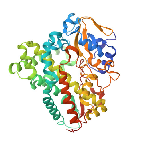
    6A7I, 6A7J

  • PubMed Abstract: 

    Bacterial cytochrome P450 (CYP) enzymes are involved in the hydroxylation of various endogenous substrates while using a heme molecule as a cofactor. CYPs have gained biotechnological interest as useful biocatalysts capable of altering chemical structures by adding a hydroxyl group in a regiospecific manner. Here, we identified, purified, and characterized two CYP154C4 proteins from Streptomyces sp. W2061 (StCYP154C4-1) and Streptomyces sp. ATCC 11861 (StCYP154C4-2). Activity assays showed that both StCYP154C4-1 and StCYP154C4-2 can produce 2'-hydroxylated testosterone, which differs from the activity of a previously described NfCYP154C5 from Nocardia farcinica in terms of its 16α-hydroxylation of testosterone. To better understand the molecular basis of the regioselectivity of these two CYP154C4 proteins, crystal structures of the ligand-unbound form of StCYP154C4-1 and the testosterone-bound form of StCYP154C4-2 were determined. Comparison with the previously determined NfCYP154C5 structure revealed differences in the substrate-binding residues, suggesting a likely explanation for the different patterns of testosterone hydroxylation, despite the high sequence similarities between the enzymes (54% identity). These findings provide valuable insights that will enable protein engineering for the development of artificial steroid-related CYPs exhibiting different regiospecificity.


  • Organizational Affiliation
    • Department of Life Science and Biochemical Engineering, Sunmoon University, Asansi, Korea.

Macromolecule Content 

  • Total Structure Weight: 45.73 kDa 
  • Atom Count: 3,194 
  • Modeled Residue Count: 407 
  • Deposited Residue Count: 411 
  • Unique protein chains: 1

Macromolecules

Find similar proteins by:|  3D Structure
Entity ID: 1
MoleculeChains  Sequence LengthOrganismDetailsImage
Cytochrome P450411Streptomyces violaceoruberMutation(s): 0 
Gene Names: B1H20_20375
UniProt
Find proteins for A0A1V0UEC8 (Streptomyces violaceoruber)
Explore A0A1V0UEC8 
Go to UniProtKB:  A0A1V0UEC8
Entity Groups
Sequence Clusters30% Identity50% Identity70% Identity90% Identity95% Identity100% Identity
UniProt GroupA0A1V0UEC8
Sequence Annotations
Expand
Reference Sequence

Small Molecules

Experimental Data & Validation

Experimental Data

  • Method: X-RAY DIFFRACTION
  • Resolution: 2.71 Å
  • R-Value Free:  0.266 (Depositor), 0.268 (DCC) 
  • R-Value Work:  0.217 (Depositor), 0.219 (DCC) 
  • R-Value Observed: 0.219 (Depositor) 
Space Group: I 21 21 21
Unit Cell:
Length ( Å )Angle ( ˚ )
a = 90.381α = 90
b = 113.94β = 90
c = 128.361γ = 90
Software Package:
Software NamePurpose
REFMACrefinement
MxDCdata collection
HKL-2000data scaling

Structure Validation

View Full Validation Report


Ligand Structure Quality Assessment 


Entry History 

Deposition Data

Revision History  (Full details and data files)

  • Version 1.0: 2019-01-16
    Type: Initial release
  • Version 1.1: 2019-05-22
    Changes: Data collection, Database references
  • Version 1.2: 2024-03-27
    Changes: Data collection, Database references