6A1K | pdb_00006a1k

Phosphate acyltransferase PlsX from B.subtilis


Experimental Data Snapshot

  • Method: X-RAY DIFFRACTION
  • Resolution: 2.30 Å
  • R-Value Free: 
    0.238 (Depositor), 0.238 (DCC) 
  • R-Value Work: 
    0.202 (Depositor), 0.204 (DCC) 
  • R-Value Observed: 
    0.204 (Depositor) 

wwPDB Validation 3D Report Full Report

Validation slider image for 6A1K

This is version 1.2 of the entry. See complete history

Literature

Identification of an amphipathic peptide sensor of the Bacillus subtilisfluid membrane microdomains.

Jiang, Y.Dai, X.Qin, M.Guo, Z.

(2019) Commun Biol 2: 316-316

  • DOI: https://doi.org/10.1038/s42003-019-0562-8
  • Primary Citation Related Structures: 
    6A1K

  • PubMed Abstract: 

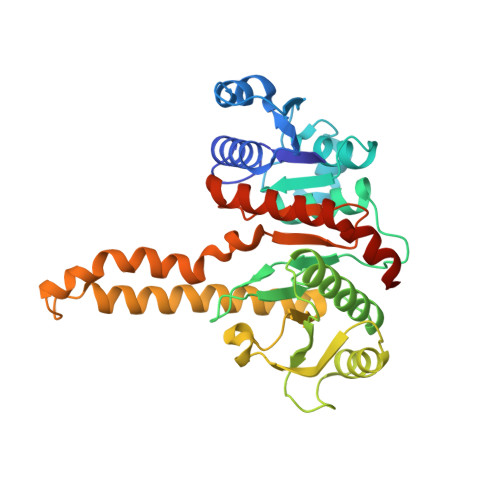
    Regions of increased fluidity are newly found bacterial membrane microdomains that are composed of short, unsaturated and branched fatty acyl chains in a fluid and disordered state. Currently, little is known about how proteins are recruited and localized to these membrane domains. Here, we identify a short amphipathic α-peptide in a previously unreported crystal structure and show that it is responsible for peripheral localization of the phosphate acyltransferase PlsX to the fluid microdomains in Bacillus subtilis . Mutations disrupting the amphipathic interaction or increasing the nonpolar interaction are found to redistribute the protein to the cytosol or other part of the plasma membrane, causing growth defects. These results reveal a mechanism of peripheral membrane sensing through optimizing nonpolar interaction with the special lipids in the microdomains. This finding shows that the fluid membrane microdomains may take advantage of their unique lipid environment as a means of recruiting and organizing proteins.


  • Organizational Affiliation
    • Shenzhen Research Institute and Department of Chemistry, The Hong Kong University of Science and Technology, Clear Water Bay, Kowloon, Hong Kong SAR China.

Macromolecule Content 

  • Total Structure Weight: 72.22 kDa 
  • Atom Count: 4,888 
  • Modeled Residue Count: 651 
  • Deposited Residue Count: 668 
  • Unique protein chains: 1

Macromolecules

Find similar proteins by:|  3D Structure
Entity ID: 1
MoleculeChains  Sequence LengthOrganismDetailsImage
Phosphate acyltransferase
A, B
334Bacillus subtilis subsp. subtilis str. 168Mutation(s): 0 
Gene Names: plsXylpDBSU15890
EC: 2.3.1 (PDB Primary Data), 2.3.1.274 (UniProt)
UniProt
Find proteins for P71018 (Bacillus subtilis (strain 168))
Explore P71018 
Go to UniProtKB:  P71018
Entity Groups
Sequence Clusters30% Identity50% Identity70% Identity90% Identity95% Identity100% Identity
UniProt GroupP71018
Sequence Annotations
Expand
Reference Sequence

Experimental Data & Validation

Experimental Data

  • Method: X-RAY DIFFRACTION
  • Resolution: 2.30 Å
  • R-Value Free:  0.238 (Depositor), 0.238 (DCC) 
  • R-Value Work:  0.202 (Depositor), 0.204 (DCC) 
  • R-Value Observed: 0.204 (Depositor) 
Space Group: C 2 2 2
Unit Cell:
Length ( Å )Angle ( ˚ )
a = 108.52α = 90
b = 144.76β = 90
c = 84.84γ = 90
Software Package:
Software NamePurpose
Aimlessdata scaling
PHENIXrefinement
PDB_EXTRACTdata extraction
MOSFLMdata reduction
PHENIXphasing

Structure Validation

View Full Validation Report


Entry History 

& Funding Information

Deposition Data


Funding OrganizationLocationGrant Number
Other governmentHong KongHKUST_621/13

Revision History  (Full details and data files)

  • Version 1.0: 2019-06-12
    Type: Initial release
  • Version 1.1: 2019-12-25
    Changes: Database references
  • Version 1.2: 2024-03-27
    Changes: Data collection, Database references, Refinement description