5ZX1 | pdb_00005zx1

Crystal structure of ENT domain from T. brucei


Experimental Data Snapshot

  • Method: X-RAY DIFFRACTION
  • Resolution: 2.31 Å
  • R-Value Free: 
    0.279 (Depositor), 0.283 (DCC) 
  • R-Value Work: 
    0.234 (Depositor), 0.249 (DCC) 

Starting Model: experimental
View more details

wwPDB Validation 3D Report Full Report

Validation slider image for 5ZX1

This is version 1.2 of the entry. See complete history

Literature

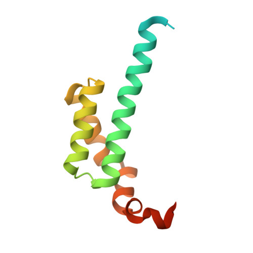
Crystal structure of an ENT domain from Trypanosoma brucei.

Mi, J.Yang, X.Zhang, J.Zhang, X.Xu, C.Liao, S.Tu, X.

(2018) Biochem Biophys Res Commun 505: 755-760

  • DOI: https://doi.org/10.1016/j.bbrc.2018.09.167
  • Primary Citation Related Structures: 
    5ZX1

  • PubMed Abstract: 

    Trypanosoma brucei (T. brucei) is a parasitic protozoan causing human sleeping sickness and related animal diseases. ENT (EMSY N-terminal) domain was first found in EMSY protein which has been proved to be involved in multiple biological processes such as DNA repair, tumorigenesis, and transcriptional regulation. So far, little is known about the function and structure of ENT domains from protozoan. Q385P5 from T. brucei, containing an ENT domain at its N-terminus, is a conserved protein in related kinetoplastid parasites. In this work, the crystal structure of ENT domain of Q385P5 (TbENT) was solved at a resolution of 2.3 Å. TbENT adopts a club-like shape consisting of five helixes, which is similar to the structure of human EMSY ENT domain (HsENT). Interestingly, TbENT shows significantly different orientation on the fifth α-helix compared with HsENT. Meanwhile, human HP1 interacts with a conserved motif adjacent to EMSY ENT domain. However, this conserved binding motif is absent in Q385P5. These differences may imply the different protein interactions and roles of Q385P5 and its ENT domain in T. brucei.


  • Organizational Affiliation
    • Hefei National Laboratory for Physical Science at Microscale and School of Life Science, University of Science and Technology of China, Hefei, Anhui, 230026, PR China.

Macromolecule Content 

  • Total Structure Weight: 13.07 kDa 
  • Atom Count: 715 
  • Modeled Residue Count: 84 
  • Deposited Residue Count: 117 
  • Unique protein chains: 1

Macromolecules

Find similar proteins by:|  3D Structure
Entity ID: 1
MoleculeChains  Sequence LengthOrganismDetailsImage
ENT117Trypanosoma brucei brucei TREU927Mutation(s): 0 
Gene Names: Tb11.02.2890
UniProt
Find proteins for Q385P5 (Trypanosoma brucei brucei (strain 927/4 GUTat10.1))
Explore Q385P5 
Go to UniProtKB:  Q385P5
Entity Groups
Sequence Clusters30% Identity50% Identity70% Identity90% Identity95% Identity100% Identity
UniProt GroupQ385P5
Sequence Annotations
Expand
Reference Sequence

Experimental Data & Validation

Experimental Data

  • Method: X-RAY DIFFRACTION
  • Resolution: 2.31 Å
  • R-Value Free:  0.279 (Depositor), 0.283 (DCC) 
  • R-Value Work:  0.234 (Depositor), 0.249 (DCC) 
Space Group: I 41 2 2
Unit Cell:
Length ( Å )Angle ( ˚ )
a = 132.59α = 90
b = 132.59β = 90
c = 40.03γ = 90
Software Package:
Software NamePurpose
PHENIXrefinement
xia2data reduction
xia2data scaling
PHENIXphasing

Structure Validation

View Full Validation Report


Entry History 

& Funding Information

Deposition Data


Funding OrganizationLocationGrant Number
National Natural Science Foundation of ChinaChina31500601

Revision History  (Full details and data files)

  • Version 1.0: 2019-05-22
    Type: Initial release
  • Version 1.1: 2020-12-02
    Changes: Database references
  • Version 1.2: 2023-11-22
    Changes: Data collection, Database references, Refinement description