5ZE8 | pdb_00005ze8

Crystal structure of a penta-heme cytochrome c552 from Thermochromatium tepidum


Experimental Data Snapshot

  • Method: X-RAY DIFFRACTION
  • Resolution: 1.60 Å
  • R-Value Free: 
    0.191 (Depositor), 0.199 (DCC) 
  • R-Value Work: 
    0.158 (Depositor), 0.170 (DCC) 
  • R-Value Observed: 
    0.160 (Depositor) 

wwPDB Validation 3D Report Full Report

Validation slider image for 5ZE8

Ligand Structure Quality Assessment 


This is version 2.1 of the entry. See complete history

Literature

Properties and structure of a low-potential, penta-heme cytochrome c552from a thermophilic purple sulfur photosynthetic bacterium Thermochromatium tepidum.

Chen, J.H.Yu, L.J.Boussac, A.Wang-Otomo, Z.Y.Kuang, T.Shen, J.R.

(2019) Photosynth Res 139: 281-293

  • DOI: https://doi.org/10.1007/s11120-018-0507-y
  • Primary Citation Related Structures: 
    5ZE8

  • PubMed Abstract: 

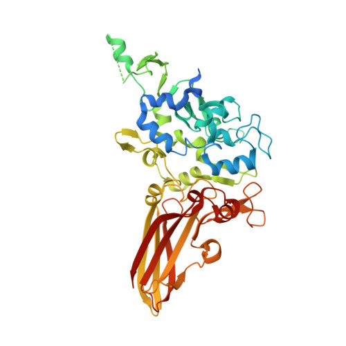
    The thermophilic purple sulfur bacterium Thermochromatium tepidum possesses four main water-soluble redox proteins involved in the electron transfer behavior. Crystal structures have been reported for three of them: a high potential iron-sulfur protein, cytochrome c', and one of two low-potential cytochrome c 552 (which is a flavocytochrome c) have been determined. In this study, we purified another low-potential cytochrome c 552 (LPC), determined its N-terminal amino acid sequence and the whole gene sequence, characterized it with absorption and electron paramagnetic spectroscopy, and solved its high-resolution crystal structure. This novel cytochrome was found to contain five c-type hemes. The overall fold of LPC consists of two distinct domains, one is the five heme-containing domain and the other one is an Ig-like domain. This provides a representative example for the structures of multiheme cytochromes containing an odd number of hemes, although the structures of multiheme cytochromes with an even number of hemes are frequently seen in the PDB database. Comparison of the sequence and structure of LPC with other proteins in the databases revealed several characteristic features which may be important for its functioning. Based on the results obtained, we discuss the possible intracellular function of this LPC in Tch. tepidum.


  • Organizational Affiliation
    • Photosynthesis Research Center, Key Laboratory of Photobiology, Institute of Botany, Chinese Academy of Sciences, No. 20, Nanxincun, Xiangshan, Beijing, 100093, China.

Macromolecule Content 

  • Total Structure Weight: 50.93 kDa 
  • Atom Count: 3,560 
  • Modeled Residue Count: 385 
  • Deposited Residue Count: 435 
  • Unique protein chains: 1

Macromolecules

Find similar proteins by:|  3D Structure
Entity ID: 1
MoleculeChains  Sequence LengthOrganismDetailsImage
cytochrome c552435Thermochromatium tepidumMutation(s): 0 
UniProt
Find proteins for A0A2P1N6U9 (Thermochromatium tepidum)
Explore A0A2P1N6U9 
Go to UniProtKB:  A0A2P1N6U9
Entity Groups
Sequence Clusters30% Identity50% Identity70% Identity90% Identity95% Identity100% Identity
UniProt GroupA0A2P1N6U9
Sequence Annotations
Expand
Reference Sequence

Small Molecules

Ligands 3 Unique
IDChains Name / Formula / InChI Key2D Diagram3D Interactions
HEC

Query on HEC


Download:Ideal Coordinates CCD File
B [auth A],
C [auth A],
D [auth A],
E [auth A],
F [auth A]
HEME C
C34 H34 Fe N4 O4
HXQIYSLZKNYNMH-LJNAALQVSA-N
SO4

Query on SO4


Download:Ideal Coordinates CCD File
G [auth A]SULFATE ION
O4 S
QAOWNCQODCNURD-UHFFFAOYSA-L
GOL

Query on GOL


Download:Ideal Coordinates CCD File
H [auth A]GLYCEROL
C3 H8 O3
PEDCQBHIVMGVHV-UHFFFAOYSA-N

Experimental Data & Validation

Experimental Data

  • Method: X-RAY DIFFRACTION
  • Resolution: 1.60 Å
  • R-Value Free:  0.191 (Depositor), 0.199 (DCC) 
  • R-Value Work:  0.158 (Depositor), 0.170 (DCC) 
  • R-Value Observed: 0.160 (Depositor) 
Space Group: C 1 2 1
Unit Cell:
Length ( Å )Angle ( ˚ )
a = 144.7α = 90
b = 39.1β = 109.31
c = 68.65γ = 90
Software Package:
Software NamePurpose
REFMACrefinement
PDB_EXTRACTdata extraction
XDSdata reduction
XDSdata scaling
SHELXDEphasing

Structure Validation

View Full Validation Report


Ligand Structure Quality Assessment 


Entry History 

Deposition Data

Revision History  (Full details and data files)

  • Version 1.0: 2018-04-25
    Type: Initial release
  • Version 1.1: 2018-05-16
    Changes: Data collection, Database references
  • Version 1.2: 2019-02-20
    Changes: Data collection, Database references
  • Version 2.0: 2019-10-02
    Changes: Atomic model, Data collection, Derived calculations, Non-polymer description, Structure summary
  • Version 2.1: 2024-10-16
    Changes: Data collection, Database references, Derived calculations, Structure summary