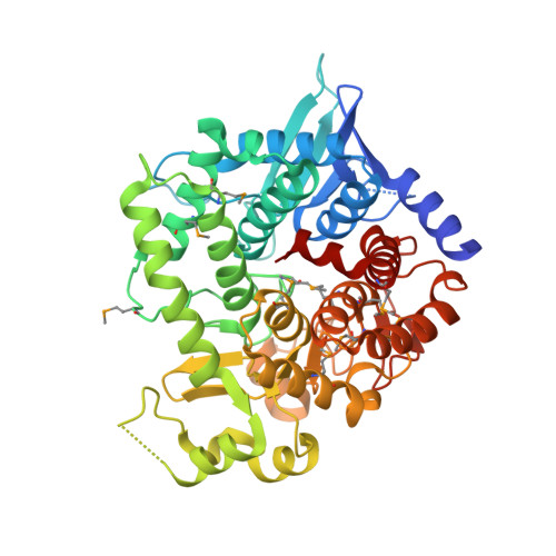
5Z74

Crystal structure of alkaline/neutral invertase InvB from Anabaena sp. PCC 7120 complexed with sucrose


Experimental Data Snapshot

  • Method: X-RAY DIFFRACTION
  • Resolution: 1.95 Å
  • R-Value Free: 0.239 
  • R-Value Work: 0.208 
  • R-Value Observed: 0.210 

Starting Model: experimental
View more details

wwPDB Validation   3D Report Full Report


This is version 2.1 of the entry. See complete history


Literature

Structural and enzymatic analyses of Anabaena heterocyst-specific alkaline invertase InvB.

Xie, J.Hu, H.X.Cai, K.Xia, L.Y.Yang, F.Jiang, Y.L.Chen, Y.Zhou, C.Z.

(2018) FEBS Lett 592: 1589-1601

  • DOI: https://doi.org/10.1002/1873-3468.13041
  • Primary Citation of Related Structures:  
    5Z73, 5Z74

  • PubMed Abstract: 

    Anabaena sp. PCC 7120 encodes two alkaline/neutral invertases, namely InvA and InvB. Following our recently reported InvA structure, here we report the crystal structure of the heterocyst-specific InvB. Despite sharing an overall structure similar to InvA, InvB possesses a much higher catalytic activity. Structural comparisons of the catalytic pockets reveal that Arg430 of InvB adopts a different conformation, which may facilitate the deprotonation of the catalytic residue Glu415. We propose that the higher activity may be responsible for the vital role of InvB in heterocyst development and nitrogen fixation. Furthermore, phylogenetic analysis combined with activity assays also suggests the role of this highly conserved arginine in plants and cyanobacteria, as well as some proteobacteria living in highly extreme environments.


  • Organizational Affiliation

    Hefei National Laboratory for Physical Sciences at the Microscale and School of Life Sciences, University of Science and Technology of China, Hefei Anhui, China.


Macromolecules
Find similar proteins by:  (by identity cutoff)  |  3D Structure
Entity ID: 1
MoleculeChains Sequence LengthOrganismDetailsImage
Alr0819 proteinA [auth B],
B [auth A]
457Nostoc sp. PCC 7120 = FACHB-418Mutation(s): 0 
Gene Names: invBalr0819
EC: 3.2.1.26
UniProt
Find proteins for Q8YYM9 (Nostoc sp. (strain PCC 7120 / SAG 25.82 / UTEX 2576))
Explore Q8YYM9 
Go to UniProtKB:  Q8YYM9
Entity Groups  
Sequence Clusters30% Identity50% Identity70% Identity90% Identity95% Identity100% Identity
UniProt GroupQ8YYM9
Sequence Annotations
Expand
  • Reference Sequence
Oligosaccharides

Help

Entity ID: 2
MoleculeChains Length2D Diagram Glycosylation3D Interactions
beta-D-fructofuranose-(2-1)-alpha-D-glucopyranose
C, D
2N/A
Glycosylation Resources
GlyTouCan:  G05551OP
GlyCosmos:  G05551OP
Small Molecules
Modified Residues  1 Unique
IDChains TypeFormula2D DiagramParent
MSE
Query on MSE
A [auth B],
B [auth A]
L-PEPTIDE LINKINGC5 H11 N O2 SeMET
Biologically Interesting Molecules (External Reference) 1 Unique
Entity ID: 2
IDChains NameType/Class2D Diagram3D Interactions
PRD_900003
Query on PRD_900003
C, D
sucroseOligosaccharide / Nutrient
Experimental Data & Validation

Experimental Data

  • Method: X-RAY DIFFRACTION
  • Resolution: 1.95 Å
  • R-Value Free: 0.239 
  • R-Value Work: 0.208 
  • R-Value Observed: 0.210 
  • Space Group: C 1 2 1
Unit Cell:
Length ( Å )Angle ( ˚ )
a = 166.946α = 90
b = 82.171β = 119.13
c = 97.446γ = 90
Software Package:
Software NamePurpose
REFMACrefinement
HKL-2000data collection

Structure Validation

View Full Validation Report


Entry History & Funding Information

Deposition Data


Funding OrganizationLocationGrant Number
National Natural Science Foundation of ChinaChina31630001

Revision History  (Full details and data files)

  • Version 1.0: 2018-05-30
    Type: Initial release
  • Version 2.0: 2020-07-29
    Type: Remediation
    Reason: Carbohydrate remediation
    Changes: Atomic model, Author supporting evidence, Data collection, Derived calculations, Non-polymer description, Structure summary
  • Version 2.1: 2023-11-22
    Changes: Data collection, Database references, Refinement description, Structure summary