5YMT | pdb_00005ymt

Functional and structural characterization of P[19] rotavirus VP8* interaction with histo-blood group antigens


Experimental Data Snapshot

  • Method: X-RAY DIFFRACTION
  • Resolution: 2.20 Å
  • R-Value Free: 
    0.267 (Depositor), 0.267 (DCC) 
  • R-Value Work: 
    0.226 (Depositor), 0.227 (DCC) 
  • R-Value Observed: 
    0.228 (Depositor) 

Starting Model: experimental
View more details

wwPDB Validation 3D Report Full Report

Validation slider image for 5YMT

This is version 2.1 of the entry. See complete history

Literature

Glycan Binding Specificity and Mechanism of Human and Porcine P[6]/P[19] Rotavirus VP8*s.

Sun, X.Li, D.Qi, J.Chai, W.Wang, L.Wang, L.Peng, R.Wang, H.Zhang, Q.Pang, L.Kong, X.Wang, H.Jin, M.Gao, G.F.Duan, Z.

(2018) J Virol 92

  • DOI: https://doi.org/10.1128/JVI.00538-18
  • Primary Citation Related Structures: 
    5YMS, 5YMT, 5YMU

  • PubMed Abstract: 

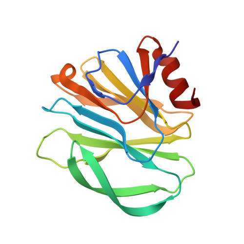
    Rotaviruses (RVs), which cause severe gastroenteritis in infants and children, recognize glycan ligands in a genotype-dependent manner via the distal VP8* head of the spike protein VP4. However, the glycan binding mechanisms remain elusive for the P[II] genogroup RVs, including the widely prevalent human RVs (P[8], P[4], and P[6]) and a rare P[19] RV. In this study, we characterized the glycan binding specificities of human and porcine P[6]/P[19] RV VP8*s and found that the P[II] genogroup RV VP8*s could commonly interact with mucin core 2, which may play an important role in RV evolution and cross-species transmission. We determined the first P[6] VP8* structure, as well as the complex structures of human P[19] VP8*, with core 2 and lacto- N -tetraose (LNT). A glycan binding site was identified in human P[19] VP8*. Structural superimposition and sequence alignment revealed the conservation of the glycan binding site in the P[II] genogroup RV VP8*s. Our data provide significant insight into the glycan binding specificity and glycan binding mechanism of the P[II] genogroup RV VP8*s, which could help in understanding RV evolution, transmission, and epidemiology and in vaccine development. IMPORTANCE Rotaviruses (RVs), belonging to the family Reoviridae , are double-stranded RNA viruses that cause acute gastroenteritis in children and animals worldwide. Depending on the phylogeny of the VP8* sequences, P[6] and P[19] RVs are grouped into genogroup II, together with P[4] and P[8], which are widely prevalent in humans. In this study, we characterized the glycan binding specificities of human and porcine P[6]/P[19] RV VP8*s, determined the crystal structure of P[6] VP8*, and uncovered the glycan binding pattern in P[19] VP8*, revealing a conserved glycan binding site in the VP8*s of P[II] genogroup RVs by structural superimposition and sequence alignment. Our data suggested that mucin core 2 may play an important role in P[II] RV evolution and cross-species transmission. These data provide insight into the cell attachment, infection, epidemiology, and evolution of P[II] genogroup RVs, which could help in developing control and prevention strategies against RVs.


  • Organizational Affiliation
    • Key Laboratory of Medical Virology and Viral Diseases, Ministry of Health of the People's Republic of China, Beijing, China.

Macromolecule Content 

  • Total Structure Weight: 255.83 kDa 
  • Atom Count: 18,722 
  • Modeled Residue Count: 2,240 
  • Deposited Residue Count: 2,240 
  • Unique protein chains: 1

Macromolecules

Find similar proteins by:|  3D Structure
Entity ID: 1
MoleculeChains  Sequence LengthOrganismDetailsImage
Outer capsid protein VP4
A, B, C, D, E
A, B, C, D, E, F, G, H, I, J, K, L, M, N
160Human rotavirus AMutation(s): 0 
UniProt
Find proteins for Q9Q2P6 (Human rotavirus A)
Explore Q9Q2P6 
Go to UniProtKB:  Q9Q2P6
Entity Groups
Sequence Clusters30% Identity50% Identity70% Identity90% Identity95% Identity100% Identity
UniProt GroupQ9Q2P6
Sequence Annotations
Expand
Reference Sequence

Oligosaccharides

Help  
Entity ID: 2
MoleculeChains Length2D Diagram GlycosylationD Interactions
beta-D-galactopyranose-(1-3)-2-acetamido-2-deoxy-beta-D-glucopyranose-(1-3)-beta-D-galactopyranose-(1-4)-alpha-D-glucopyranose
O, P, Q
4N/A
Glycosylation Resources
GlyTouCan: G19979XJ
GlyCosmos: G19979XJ
GlyGen: G19979XJ

Experimental Data & Validation

Experimental Data

  • Method: X-RAY DIFFRACTION
  • Resolution: 2.20 Å
  • R-Value Free:  0.267 (Depositor), 0.267 (DCC) 
  • R-Value Work:  0.226 (Depositor), 0.227 (DCC) 
  • R-Value Observed: 0.228 (Depositor) 
Space Group: P 32
Unit Cell:
Length ( Å )Angle ( ˚ )
a = 131.667α = 90
b = 131.667β = 90
c = 150.53γ = 120
Software Package:
Software NamePurpose
SCALEPACKdata scaling
PHENIXrefinement
PDB_EXTRACTdata extraction
HKL-2000data reduction
PHENIXphasing

Structure Validation

View Full Validation Report


Entry History 

& Funding Information

Deposition Data


Funding OrganizationLocationGrant Number
National Natural Science Foundation of ChinaChina81472003; 31500139;81601813

Revision History  (Full details and data files)

  • Version 1.0: 2018-05-02
    Type: Initial release
  • Version 1.1: 2018-05-23
    Changes: Data collection, Database references
  • Version 1.2: 2018-07-11
    Changes: Data collection, Database references
  • Version 2.0: 2020-07-29
    Type: Remediation
    Reason: Carbohydrate remediation
    Changes: Advisory, Atomic model, Data collection, Derived calculations, Structure summary
  • Version 2.1: 2023-11-22
    Changes: Data collection, Database references, Derived calculations, Refinement description, Structure summary