5YD2 | pdb_00005yd2

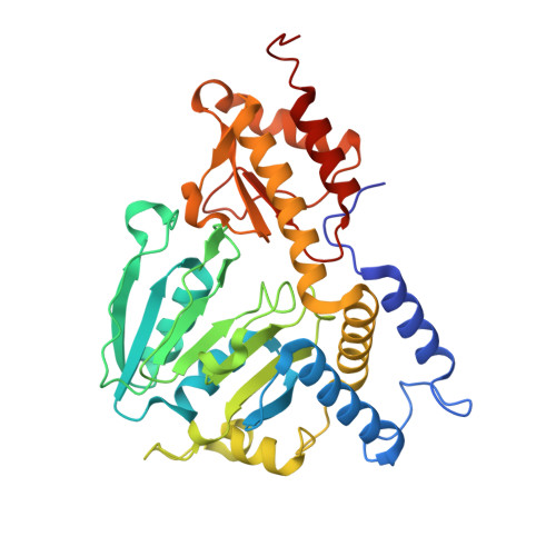
Crystal Structure of Delta 4 mutant of EhPSAT (Phosphoserine aminotransferase of Entamoeba histolytica)


Experimental Data Snapshot

  • Method: X-RAY DIFFRACTION
  • Resolution: 2.35 Å
  • R-Value Free: 
    0.287 (Depositor), 0.282 (DCC) 
  • R-Value Work: 
    0.229 (Depositor), 0.229 (DCC) 
  • R-Value Observed: 
    0.232 (Depositor) 

Starting Model: experimental
View more details

wwPDB Validation 3D Report Full Report

Validation slider image for 5YD2

Ligand Structure Quality Assessment 


This is version 1.2 of the entry. See complete history. 

Literature

N-terminal residues are crucial for quaternary structure and active site conformation for the phosphoserine aminotransferase from enteric human parasite E. histolytica.

Singh, R.K., Tomar, P., Dharavath, S., Kumar, S., Gourinath, S.

(2019) Int J Biol Macromol 132: 1012-1023

  • DOI: https://doi.org/10.1016/j.ijbiomac.2019.04.027
  • Primary Citation Related Structures: 
    5YB0, 5YD2, 5YII

  • PubMed Abstract: 

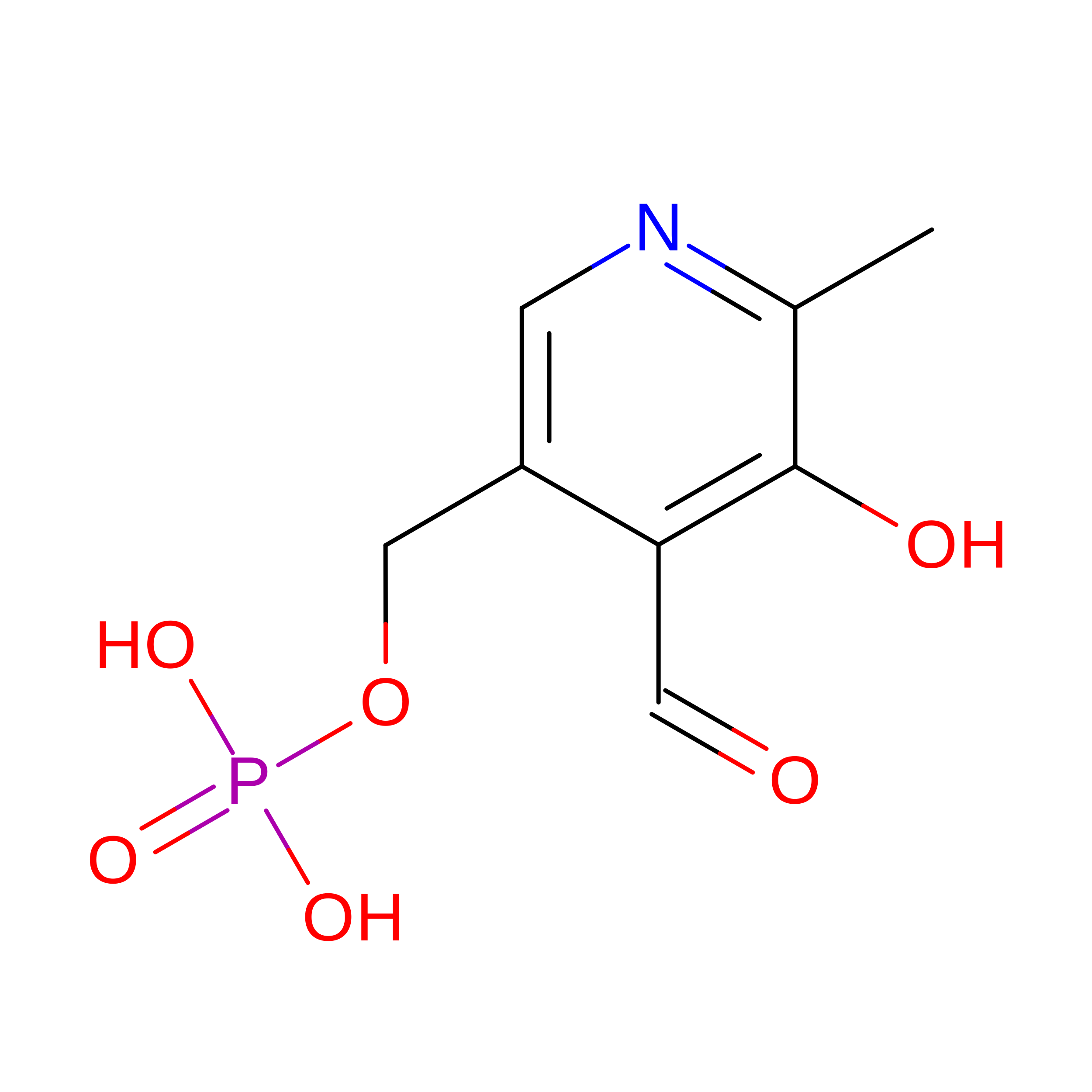
    Phosphoserine aminotransferase (PSAT) is a pyridoxal-5'phosphate (PLP)-dependent enzyme that catalyzes the second reversible step in the phosphoserine biosynthetic pathway producing serine. The crystal structure of E. histolytica PSAT (EhPSAT) complexed with PLP was elucidated at 3.0 Å resolution and the structures of its mutants, EhPSAT_Δ45 and EhPSAT_Δ4, at 1.8 and 2.4 Å resolution respectively. Deletion of 45 N-terminal residues (EhPSAT_Δ45) resulted in an inactive protein, the structure showed a dimeric arrangement drastically different from that of the wild-type protein, with the two monomers translated and rotated by almost 180° with respect to each other; causing a rearrangement of the active site to which PLP was unable to bind. Deletion of first N-terminal 15 (EhPSAT_Δ15) and four 11th to 14th residues (EhPSAT_Δ4) yielded up to 98% and 90% decrease in the activity respectively. Absence of aldimine linkage between PLP-Lys in the crystal structure of EhPSAT_Δ4 mutant explains for such decrease in activity and describes the importance of these N-terminal residues. Furthermore, a halide-binding site was found in close proximity to the active site. A stretch of six amino acids (146-NNTIYG-151) only conserved in the Entamoeba genus, contributes to halide binding may explain that the halide inhibition could be specific to Entamoeba.


  • Organizational Affiliation: 
    • Structural Biology Laboratory, School of Life Sciences, Jawaharlal Nehru University, New Delhi 110067, India.

Macromolecule Content 

  • Total Structure Weight: 81.14 kDa 
  • Atom Count: 5,597 
  • Modeled Residue Count: 695 
  • Deposited Residue Count: 708 
  • Unique protein chains: 1

Macromolecules

Find similar proteins by:|  3D Structure
Entity ID: 1
MoleculeChains  Sequence LengthOrganismDetailsImage
Phosphoserine aminotransferase
A, B
354Entamoeba histolytica HM-3:IMSSMutation(s): 0 
Gene Names: KM1_040390
EC: 2.6.1.52
UniProt
Find proteins for M7XC02 (Entamoeba histolytica HM-3:IMSS)
Explore M7XC02 
Go to UniProtKB:  M7XC02
Entity Groups
Sequence Clusters30% Identity50% Identity70% Identity90% Identity95% Identity100% Identity
UniProt GroupM7XC02
Sequence Annotations
Expand
Reference Sequence

Experimental Data & Validation

Experimental Data

  • Method: X-RAY DIFFRACTION
  • Resolution: 2.35 Å
  • R-Value Free:  0.287 (Depositor), 0.282 (DCC) 
  • R-Value Work:  0.229 (Depositor), 0.229 (DCC) 
  • R-Value Observed: 0.232 (Depositor) 
Space Group: C 1 2 1
Unit Cell:
Length ( Å )Angle ( ˚ )
a = 132.238α = 90
b = 67.904β = 110.93
c = 95.03γ = 90
Software Package:
Software NamePurpose
REFMACrefinement
HKL-2000data collection
HKL-2000data scaling
HKL-2000data reduction
DMphasing

Structure Validation

View Full Validation Report


Ligand Structure Quality Assessment 


Entry History 

Deposition Data

Revision History  (Full details and data files)

  • Version 1.0: 2018-10-31
    Type: Initial release
  • Version 1.1: 2019-07-10
    Changes: Data collection, Database references
  • Version 1.2: 2023-11-22
    Changes: Data collection, Database references, Refinement description