5XNP | pdb_00005xnp

Crystal structures of human SALM5 in complex with human PTPdelta


Experimental Data Snapshot

  • Method: X-RAY DIFFRACTION
  • Resolution: 3.73 Å
  • R-Value Free: 
    0.264 (Depositor), 0.259 (DCC) 
  • R-Value Work: 
    0.232 (Depositor), 0.230 (DCC) 
  • R-Value Observed: 
    0.233 (Depositor) 

Starting Models: experimental
View more details

wwPDB Validation 3D Report Full Report

Validation slider image for 5XNP

Ligand Structure Quality Assessment 


This is version 1.4 of the entry. See complete history

Literature

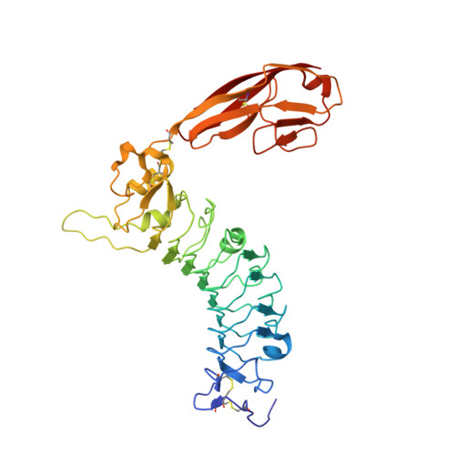
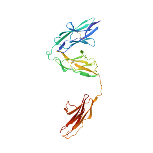
Structural basis of SALM5-induced PTP delta dimerization for synaptic differentiation

Lin, Z.Liu, J.Ding, H.Xu, F.Liu, H.

(2018) Nat Commun 9: 268-268

  • DOI: https://doi.org/10.1038/s41467-017-02414-2
  • Primary Citation Related Structures: 
    5XNP, 5XNQ

  • PubMed Abstract: 

    SALM5, a synaptic adhesion molecule implicated in autism, induces presynaptic differentiation through binding to the LAR family receptor protein tyrosine phosphatases (LAR-RPTPs) that have been highlighted as presynaptic hubs for synapse formation. The mechanisms underlying SALM5/LAR-RPTP interaction remain unsolved. Here we report crystal structures of human SALM5 LRR-Ig alone and in complex with human PTPδ Ig1-3 (MeA - ). Distinct from other LAR-RPTP ligands, SALM5 mainly exists as a dimer with LRR domains from two protomers packed in an antiparallel fashion. In the 2:2 heterotetrameric SALM5/PTPδ complex, a SALM5 dimer bridges two separate PTPδ molecules. Structure-guided mutations and heterologous synapse formation assays demonstrate that dimerization of SALM5 is prerequisite for its functionality in inducing synaptic differentiation. This study presents a structural template for the SALM family and reveals a mechanism for how a synaptic adhesion molecule directly induces cis-dimerization of LAR-RPTPs into higher-order signaling assembly.


  • Organizational Affiliation
    • State Key Laboratory of Natural and Biomimetic Drugs & Department of Molecular and Cellular Pharmacology, School of Pharmaceutical Sciences, Peking University Health Science Center, 38 Xueyuan Road, Haidian District, Beijing, 100191, China.

Macromolecule Content 

  • Total Structure Weight: 148.14 kDa 
  • Atom Count: 10,418 
  • Modeled Residue Count: 1,304 
  • Deposited Residue Count: 1,304 
  • Unique protein chains: 2

Macromolecules

Find similar proteins by:|  3D Structure
Entity ID: 1
MoleculeChains  Sequence LengthOrganismDetailsImage
Leucine-rich repeat and fibronectin type-III domain-containing protein 5
A, B
361Homo sapiensMutation(s): 0 
Gene Names: LRFN5C14orf146SALM5
UniProt & NIH Common Fund Data Resources
Find proteins for Q96NI6 (Homo sapiens)
Explore Q96NI6 
Go to UniProtKB:  Q96NI6
PHAROS:  Q96NI6
GTEx:  ENSG00000165379 
Entity Groups
Sequence Clusters30% Identity50% Identity70% Identity90% Identity95% Identity100% Identity
UniProt GroupQ96NI6
Glycosylation
Glycosylation Sites: 3Go to GlyGen: Q96NI6-1
Sequence Annotations
Expand
Reference Sequence
Find similar proteins by:|  3D Structure
Entity ID: 2
MoleculeChains  Sequence LengthOrganismDetailsImage
Receptor-type tyrosine-protein phosphatase deltaC [auth D],
D [auth E]
291Homo sapiensMutation(s): 0 
Gene Names: PTPRD
EC: 3.1.3.48
UniProt & NIH Common Fund Data Resources
Find proteins for P23468 (Homo sapiens)
Explore P23468 
Go to UniProtKB:  P23468
PHAROS:  P23468
GTEx:  ENSG00000153707 
Entity Groups
Sequence Clusters30% Identity50% Identity70% Identity90% Identity95% Identity100% Identity
UniProt GroupP23468
Glycosylation
Glycosylation Sites: 1Go to GlyGen: P23468-1
Sequence Annotations
Expand
Reference Sequence

Small Molecules

Ligands 5 Unique
IDChains Name / Formula / InChI Key2D Diagram3D Interactions
NAG

Query on NAG


Download:Ideal Coordinates CCD File
E [auth A]
F [auth A]
G [auth A]
IA [auth D]
MA [auth E]
E [auth A],
F [auth A],
G [auth A],
IA [auth D],
MA [auth E],
T [auth B],
U [auth B],
V [auth B]
2-acetamido-2-deoxy-beta-D-glucopyranose
C8 H15 N O6
OVRNDRQMDRJTHS-FMDGEEDCSA-N
SO4

Query on SO4


Download:Ideal Coordinates CCD File
GA [auth B],
HA [auth B],
R [auth A],
S [auth A]
SULFATE ION
O4 S
QAOWNCQODCNURD-UHFFFAOYSA-L
CA

Query on CA


Download:Ideal Coordinates CCD File
AA [auth B]
BA [auth B]
CA [auth B]
I [auth A]
J [auth A]
AA [auth B],
BA [auth B],
CA [auth B],
I [auth A],
J [auth A],
K [auth A],
L [auth A],
M [auth A],
N [auth A],
X [auth B],
Y [auth B],
Z [auth B]
CALCIUM ION
Ca
BHPQYMZQTOCNFJ-UHFFFAOYSA-N
CL

Query on CL


Download:Ideal Coordinates CCD File
DA [auth B]
EA [auth B]
FA [auth B]
JA [auth D]
KA [auth D]
DA [auth B],
EA [auth B],
FA [auth B],
JA [auth D],
KA [auth D],
LA [auth D],
NA [auth E],
O [auth A],
OA [auth E],
P [auth A],
PA [auth E],
Q [auth A]
CHLORIDE ION
Cl
VEXZGXHMUGYJMC-UHFFFAOYSA-M
NA

Query on NA


Download:Ideal Coordinates CCD File
H [auth A],
W [auth B]
SODIUM ION
Na
FKNQFGJONOIPTF-UHFFFAOYSA-N

Experimental Data & Validation

Experimental Data

  • Method: X-RAY DIFFRACTION
  • Resolution: 3.73 Å
  • R-Value Free:  0.264 (Depositor), 0.259 (DCC) 
  • R-Value Work:  0.232 (Depositor), 0.230 (DCC) 
  • R-Value Observed: 0.233 (Depositor) 
Space Group: I 2 3
Unit Cell:
Length ( Å )Angle ( ˚ )
a = 249.384α = 90
b = 249.384β = 90
c = 249.384γ = 90
Software Package:
Software NamePurpose
PHENIXrefinement
HKL-3000data reduction
HKL-3000data scaling
PHASERphasing

Structure Validation

View Full Validation Report


Ligand Structure Quality Assessment 


Entry History 

Deposition Data

Revision History  (Full details and data files)

  • Version 1.0: 2018-01-24
    Type: Initial release
  • Version 1.1: 2018-02-07
    Changes: Database references
  • Version 1.2: 2020-07-29
    Type: Remediation
    Reason: Carbohydrate remediation
    Changes: Data collection, Derived calculations, Structure summary
  • Version 1.3: 2023-11-22
    Changes: Data collection, Database references, Refinement description, Structure summary
  • Version 1.4: 2024-10-30
    Changes: Structure summary