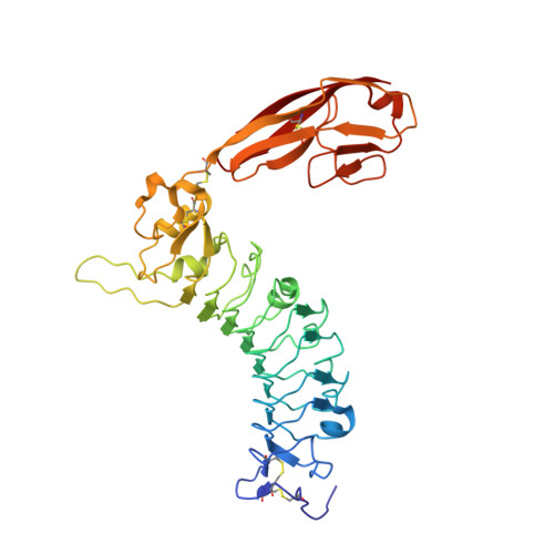
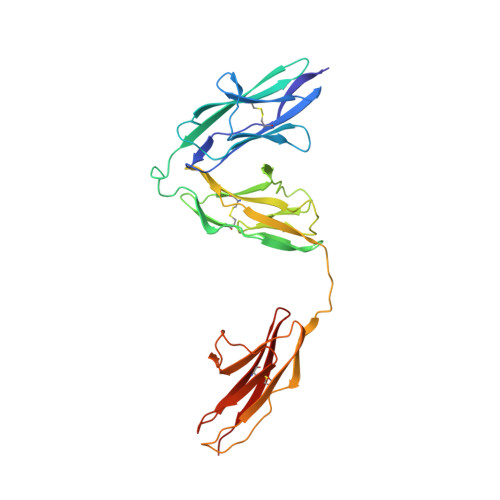
5XNP

Crystal structures of human SALM5 in complex with human PTPdelta


Experimental Data Snapshot

  • Method: X-RAY DIFFRACTION
  • Resolution: 3.73 Å
  • R-Value Free: 0.264 
  • R-Value Work: 0.232 
  • R-Value Observed: 0.233 

Starting Models: experimental
View more details

wwPDB Validation   3D Report Full Report


Ligand Structure Quality Assessment 


This is version 1.4 of the entry. See complete history


Literature

Structural basis of SALM5-induced PTP delta dimerization for synaptic differentiation

Lin, Z.Liu, J.Ding, H.Xu, F.Liu, H.

(2018) Nat Commun 9: 268-268

  • DOI: https://doi.org/10.1038/s41467-017-02414-2
  • Primary Citation of Related Structures:  
    5XNP, 5XNQ

  • PubMed Abstract: 

    SALM5, a synaptic adhesion molecule implicated in autism, induces presynaptic differentiation through binding to the LAR family receptor protein tyrosine phosphatases (LAR-RPTPs) that have been highlighted as presynaptic hubs for synapse formation. The mechanisms underlying SALM5/LAR-RPTP interaction remain unsolved. Here we report crystal structures of human SALM5 LRR-Ig alone and in complex with human PTPδ Ig1-3 (MeA - ). Distinct from other LAR-RPTP ligands, SALM5 mainly exists as a dimer with LRR domains from two protomers packed in an antiparallel fashion. In the 2:2 heterotetrameric SALM5/PTPδ complex, a SALM5 dimer bridges two separate PTPδ molecules. Structure-guided mutations and heterologous synapse formation assays demonstrate that dimerization of SALM5 is prerequisite for its functionality in inducing synaptic differentiation. This study presents a structural template for the SALM family and reveals a mechanism for how a synaptic adhesion molecule directly induces cis-dimerization of LAR-RPTPs into higher-order signaling assembly.


  • Organizational Affiliation

    State Key Laboratory of Natural and Biomimetic Drugs & Department of Molecular and Cellular Pharmacology, School of Pharmaceutical Sciences, Peking University Health Science Center, 38 Xueyuan Road, Haidian District, Beijing, 100191, China.


Macromolecules
Find similar proteins by:  (by identity cutoff)  |  3D Structure
Entity ID: 1
MoleculeChains Sequence LengthOrganismDetailsImage
Leucine-rich repeat and fibronectin type-III domain-containing protein 5
A, B
361Homo sapiensMutation(s): 0 
Gene Names: LRFN5C14orf146SALM5
UniProt & NIH Common Fund Data Resources
Find proteins for Q96NI6 (Homo sapiens)
Explore Q96NI6 
Go to UniProtKB:  Q96NI6
PHAROS:  Q96NI6
GTEx:  ENSG00000165379 
Entity Groups  
Sequence Clusters30% Identity50% Identity70% Identity90% Identity95% Identity100% Identity
UniProt GroupQ96NI6
Glycosylation
Glycosylation Sites: 3Go to GlyGen: Q96NI6-1
Sequence Annotations
Expand
  • Reference Sequence
Find similar proteins by:  (by identity cutoff)  |  3D Structure
Entity ID: 2
MoleculeChains Sequence LengthOrganismDetailsImage
Receptor-type tyrosine-protein phosphatase deltaC [auth D],
D [auth E]
291Homo sapiensMutation(s): 0 
Gene Names: PTPRD
EC: 3.1.3.48
UniProt & NIH Common Fund Data Resources
Find proteins for P23468 (Homo sapiens)
Explore P23468 
Go to UniProtKB:  P23468
PHAROS:  P23468
GTEx:  ENSG00000153707 
Entity Groups  
Sequence Clusters30% Identity50% Identity70% Identity90% Identity95% Identity100% Identity
UniProt GroupP23468
Glycosylation
Glycosylation Sites: 1Go to GlyGen: P23468-1
Sequence Annotations
Expand
  • Reference Sequence
Small Molecules
Ligands 5 Unique
IDChains Name / Formula / InChI Key2D Diagram3D Interactions
NAG
Query on NAG

Download Ideal Coordinates CCD File 
E [auth A]
F [auth A]
G [auth A]
IA [auth D]
MA [auth E]
E [auth A],
F [auth A],
G [auth A],
IA [auth D],
MA [auth E],
T [auth B],
U [auth B],
V [auth B]
2-acetamido-2-deoxy-beta-D-glucopyranose
C8 H15 N O6
OVRNDRQMDRJTHS-FMDGEEDCSA-N
SO4
Query on SO4

Download Ideal Coordinates CCD File 
GA [auth B],
HA [auth B],
R [auth A],
S [auth A]
SULFATE ION
O4 S
QAOWNCQODCNURD-UHFFFAOYSA-L
CA
Query on CA

Download Ideal Coordinates CCD File 
AA [auth B]
BA [auth B]
CA [auth B]
I [auth A]
J [auth A]
AA [auth B],
BA [auth B],
CA [auth B],
I [auth A],
J [auth A],
K [auth A],
L [auth A],
M [auth A],
N [auth A],
X [auth B],
Y [auth B],
Z [auth B]
CALCIUM ION
Ca
BHPQYMZQTOCNFJ-UHFFFAOYSA-N
CL
Query on CL

Download Ideal Coordinates CCD File 
DA [auth B]
EA [auth B]
FA [auth B]
JA [auth D]
KA [auth D]
DA [auth B],
EA [auth B],
FA [auth B],
JA [auth D],
KA [auth D],
LA [auth D],
NA [auth E],
O [auth A],
OA [auth E],
P [auth A],
PA [auth E],
Q [auth A]
CHLORIDE ION
Cl
VEXZGXHMUGYJMC-UHFFFAOYSA-M
NA
Query on NA

Download Ideal Coordinates CCD File 
H [auth A],
W [auth B]
SODIUM ION
Na
FKNQFGJONOIPTF-UHFFFAOYSA-N
Experimental Data & Validation

Experimental Data

  • Method: X-RAY DIFFRACTION
  • Resolution: 3.73 Å
  • R-Value Free: 0.264 
  • R-Value Work: 0.232 
  • R-Value Observed: 0.233 
  • Space Group: I 2 3
Unit Cell:
Length ( Å )Angle ( ˚ )
a = 249.384α = 90
b = 249.384β = 90
c = 249.384γ = 90
Software Package:
Software NamePurpose
PHENIXrefinement
HKL-3000data reduction
HKL-3000data scaling
PHASERphasing

Structure Validation

View Full Validation Report


Ligand Structure Quality Assessment 


Entry History 

Deposition Data

Revision History  (Full details and data files)

  • Version 1.0: 2018-01-24
    Type: Initial release
  • Version 1.1: 2018-02-07
    Changes: Database references
  • Version 1.2: 2020-07-29
    Type: Remediation
    Reason: Carbohydrate remediation
    Changes: Data collection, Derived calculations, Structure summary
  • Version 1.3: 2023-11-22
    Changes: Data collection, Database references, Refinement description, Structure summary
  • Version 1.4: 2024-10-30
    Changes: Structure summary