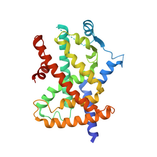
5WR1 | pdb_00005wr1

Covalent bond formation of bifunctional ligand with hPPARg-LBD


Experimental Data Snapshot

  • Method: X-RAY DIFFRACTION
  • Resolution: 2.34 Å
  • R-Value Free: 
    0.259 (Depositor), 0.260 (DCC) 
  • R-Value Work: 
    0.229 (Depositor), 0.230 (DCC) 
  • R-Value Observed: 
    0.231 (Depositor) 

wwPDB Validation   3D Report Full Report


Ligand Structure Quality Assessment 

Created with Raphaël 2.3.0Worse 01 BetterLigand structure goodness of fit to experimental dataBest fitted 8XOClick on this verticalbar to view details

This is version 1.2 of the entry. See complete history


Literature

On-site reaction for PPAR gamma modification using a specific bifunctional ligand

Kojima, H.Itoh, T.Yamamoto, K.

(2017) Bioorg Med Chem 25: 6492-6500

  • DOI: https://doi.org/10.1016/j.bmc.2017.10.024
  • Primary Citation of Related Structures:  
    5WQX, 5WR0, 5WR1

  • PubMed Abstract: 

    Site-specific labeling is an important methodology to elucidate the biological function of a target protein. Here, we report a strategy for site-specific chemical labeling, termed the "on-site reaction". We designed and readily synthesized a bifunctional ligand possessing two reaction sites, an enone and an azide moiety. This strategy involves an on-site conjugate addition reaction with protein followed by a Hüisgen cycloaddition reaction. We demonstrate this strategy by using fluorescein as a probe and peroxisome proliferator activated receptor γ (PPARγ) as a target protein. The reactions were evaluated by ESI-mass analysis and the binding site and modes of binding were revealed by X-ray crystallization analysis. The proposed methodology can easily convert a covalent ligand into chemical tool for protein functional analysis and the identification of drug targets.


  • Organizational Affiliation

    Laboratory of Drug Design and Medicinal Chemistry, Showa Pharmaceutical University, 3-3165 Higashi-Tamagawagakuen, Machida, Tokyo 194-8543, Japan.


Macromolecules
Find similar proteins by:  (by identity cutoff)  |  3D Structure
Entity ID: 1
MoleculeChains Sequence LengthOrganismDetailsImage
Peroxisome proliferator-activated receptor gamma
A, B
276Homo sapiensMutation(s): 0 
Gene Names: PPARGNR1C3
UniProt & NIH Common Fund Data Resources
Find proteins for P37231 (Homo sapiens)
Explore P37231 
Go to UniProtKB:  P37231
PHAROS:  P37231
GTEx:  ENSG00000132170 
Entity Groups  
Sequence Clusters30% Identity50% Identity70% Identity90% Identity95% Identity100% Identity
UniProt GroupP37231
Sequence Annotations
Expand
  • Reference Sequence
Small Molecules
Ligands 1 Unique
IDChains Name / Formula / InChI Key2D Diagram3D Interactions
8XO
Query on 8XO

Download Ideal Coordinates CCD File 
C [auth A]2-[E-(E-16-azido-2-oxidanylidene-hexadec-3-enylidene)amino]ethanoic acid
C18 H30 N4 O3
VMJXHBQSQBKHEB-PZBCRXGTSA-N
Experimental Data & Validation

Experimental Data

  • Method: X-RAY DIFFRACTION
  • Resolution: 2.34 Å
  • R-Value Free:  0.259 (Depositor), 0.260 (DCC) 
  • R-Value Work:  0.229 (Depositor), 0.230 (DCC) 
  • R-Value Observed: 0.231 (Depositor) 
Space Group: C 1 2 1
Unit Cell:
Length ( Å )Angle ( ˚ )
a = 93α = 90
b = 61.43β = 102.85
c = 118.5γ = 90
Software Package:
Software NamePurpose
REFMACrefinement

Structure Validation

View Full Validation Report


Ligand Structure Quality Assessment 

Created with Raphaël 2.3.0Worse 01 BetterLigand structure goodness of fit to experimental dataBest fitted 8XOClick on this verticalbar to view details

Entry History 

Deposition Data

Revision History  (Full details and data files)

  • Version 1.0: 2017-11-22
    Type: Initial release
  • Version 1.1: 2017-12-13
    Changes: Database references
  • Version 1.2: 2024-10-16
    Changes: Data collection, Database references, Structure summary