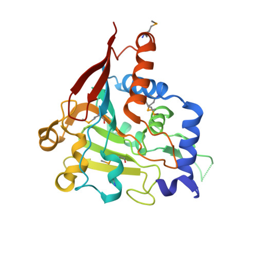
5V46

Crystal structure of the I113M, F270M, K291M, L308M mutant of SR1 domain of human sacsin


Experimental Data Snapshot

  • Method: X-RAY DIFFRACTION
  • Resolution: 1.80 Å
  • R-Value Free: 0.230 
  • R-Value Work: 0.198 
  • R-Value Observed: 0.200 

Starting Model: experimental
View more details

wwPDB Validation   3D Report Full Report


This is version 1.5 of the entry. See complete history


Literature

Structures of ubiquitin-like (Ubl) and Hsp90-like domains of sacsin provide insight into pathological mutations.

Menade, M.Kozlov, G.Trempe, J.F.Pande, H.Shenker, S.Wickremasinghe, S.Li, X.Hojjat, H.Dicaire, M.J.Brais, B.McPherson, P.S.Wong, M.J.H.Young, J.C.Gehring, K.

(2018) J Biol Chem 293: 12832-12842

  • DOI: https://doi.org/10.1074/jbc.RA118.003939
  • Primary Citation of Related Structures:  
    5V44, 5V45, 5V46, 5V47, 5VSX, 5VSZ

  • PubMed Abstract: 

    Autosomal recessive spastic ataxia of Charlevoix-Saguenay (ARSACS) is a neurodegenerative disease that is caused by mutations in the SACS gene. The product of this gene is a very large 520-kDa cytoplasmic protein, sacsin, with a ubiquitin-like (Ubl) domain at the N terminus followed by three large sacsin internal repeat (SIRPT) supradomains and C-terminal J and HEPN domains. The SIRPTs are predicted to contain Hsp90-like domains, suggesting a potential chaperone activity. In this work, we report the structures of the Hsp90-like Sr1 domain of SIRPT1 and the N-terminal Ubl domain determined at 1.55- and 2.1-Å resolutions, respectively. The Ubl domain crystallized as a swapped dimer that could be relevant in the context of full-length protein. The Sr1 domain displays the Bergerat protein fold with a characteristic nucleotide-binding pocket, although it binds nucleotides with very low affinity. The Sr1 structure reveals that ARSACS-causing missense mutations (R272H, R272C, and T201K) disrupt protein folding, most likely leading to sacsin degradation. This work lends structural support to the view of sacsin as a molecular chaperone and provides a framework for future studies of this protein.


  • Organizational Affiliation

    Department of Biochemistry, McGill Centre for Structural Biology, McGill University, Montreal, Quebec H3G 0B1, Canada.


Macromolecules
Find similar proteins by:  (by identity cutoff)  |  3D Structure
Entity ID: 1
MoleculeChains Sequence LengthOrganismDetailsImage
Sacsin
A, B
253Homo sapiensMutation(s): 4 
Gene Names: SACSKIAA0730
UniProt & NIH Common Fund Data Resources
Find proteins for Q9NZJ4 (Homo sapiens)
Explore Q9NZJ4 
Go to UniProtKB:  Q9NZJ4
PHAROS:  Q9NZJ4
GTEx:  ENSG00000151835 
Entity Groups  
Sequence Clusters30% Identity50% Identity70% Identity90% Identity95% Identity100% Identity
UniProt GroupQ9NZJ4
Sequence Annotations
Expand
  • Reference Sequence
Small Molecules
Modified Residues  1 Unique
IDChains TypeFormula2D DiagramParent
MSE
Query on MSE
A, B
L-PEPTIDE LINKINGC5 H11 N O2 SeMET
Experimental Data & Validation

Experimental Data

  • Method: X-RAY DIFFRACTION
  • Resolution: 1.80 Å
  • R-Value Free: 0.230 
  • R-Value Work: 0.198 
  • R-Value Observed: 0.200 
  • Space Group: P 43
Unit Cell:
Length ( Å )Angle ( ˚ )
a = 70.753α = 90
b = 70.753β = 90
c = 100.848γ = 90
Software Package:
Software NamePurpose
REFMACrefinement
HKL-2000data reduction
HKL-2000data scaling
PHASERphasing

Structure Validation

View Full Validation Report


Entry History & Funding Information

Deposition Data


Funding OrganizationLocationGrant Number
ARSACS FoundationCanada--

Revision History  (Full details and data files)

  • Version 1.0: 2017-05-24
    Type: Initial release
  • Version 1.1: 2018-07-11
    Changes: Data collection, Database references
  • Version 1.2: 2018-08-29
    Changes: Data collection, Database references
  • Version 1.3: 2023-10-04
    Changes: Data collection, Database references, Refinement description
  • Version 1.4: 2023-11-15
    Changes: Data collection
  • Version 1.5: 2024-11-13
    Changes: Structure summary