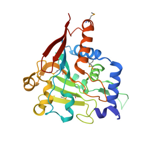
5V46 | pdb_00005v46

Crystal structure of the I113M, F270M, K291M, L308M mutant of SR1 domain of human sacsin


Experimental Data Snapshot

  • Method: X-RAY DIFFRACTION
  • Resolution: 1.80 Å
  • R-Value Free: 
    0.230 (Depositor), 0.171 (DCC) 
  • R-Value Work: 
    0.198 (Depositor) 
  • R-Value Observed: 
    0.200 (Depositor) 

Starting Model: experimental
View more details

wwPDB Validation 3D Report Full Report

Validation slider image for 5V46

This is version 1.5 of the entry. See complete history. 

Literature

Structures of ubiquitin-like (Ubl) and Hsp90-like domains of sacsin provide insight into pathological mutations.

Menade, M., Kozlov, G., Trempe, J.F., Pande, H., Shenker, S., Wickremasinghe, S., Li, X., Hojjat, H., Dicaire, M.J., Brais, B., McPherson, P.S., Wong, M.J.H., Young, J.C., Gehring, K.

(2018) J Biol Chem 293: 12832-12842

  • DOI: https://doi.org/10.1074/jbc.RA118.003939
  • Primary Citation Related Structures: 
    5V44, 5V45, 5V46, 5V47, 5VSX, 5VSZ

  • PubMed Abstract: 

    Autosomal recessive spastic ataxia of Charlevoix-Saguenay (ARSACS) is a neurodegenerative disease that is caused by mutations in the SACS gene. The product of this gene is a very large 520-kDa cytoplasmic protein, sacsin, with a ubiquitin-like (Ubl) domain at the N terminus followed by three large sacsin internal repeat (SIRPT) supradomains and C-terminal J and HEPN domains. The SIRPTs are predicted to contain Hsp90-like domains, suggesting a potential chaperone activity. In this work, we report the structures of the Hsp90-like Sr1 domain of SIRPT1 and the N-terminal Ubl domain determined at 1.55- and 2.1-Å resolutions, respectively. The Ubl domain crystallized as a swapped dimer that could be relevant in the context of full-length protein. The Sr1 domain displays the Bergerat protein fold with a characteristic nucleotide-binding pocket, although it binds nucleotides with very low affinity. The Sr1 structure reveals that ARSACS-causing missense mutations (R272H, R272C, and T201K) disrupt protein folding, most likely leading to sacsin degradation. This work lends structural support to the view of sacsin as a molecular chaperone and provides a framework for future studies of this protein.


  • Organizational Affiliation: 
    • Department of Biochemistry, McGill Centre for Structural Biology, McGill University, Montreal, Quebec H3G 0B1, Canada.

Macromolecule Content 

  • Total Structure Weight: 57.69 kDa 
  • Atom Count: 4,153 
  • Modeled Residue Count: 494 
  • Deposited Residue Count: 506 
  • Unique protein chains: 1

Macromolecules

Find similar proteins by:|  3D Structure
Entity ID: 1
MoleculeChains  Sequence LengthOrganismDetailsImage
Sacsin
A, B
253Homo sapiensMutation(s): 4 
Gene Names: SACS, KIAA0730
UniProt & NIH Common Fund Data Resources
Find proteins for Q9NZJ4 (Homo sapiens)
Explore Q9NZJ4 
Go to UniProtKB:  Q9NZJ4
PHAROS:  Q9NZJ4
GTEx:  ENSG00000151835 
Entity Groups
Sequence Clusters30% Identity50% Identity70% Identity90% Identity95% Identity100% Identity
UniProt GroupQ9NZJ4
Sequence Annotations
Expand
Reference Sequence

Small Molecules

Modified Residues  1 Unique
IDChains TypeFormula2D DiagramParent
MSE
Query on MSE
A, B
L-PEPTIDE LINKINGC5 H11 N O2 SeMET

Experimental Data & Validation

Experimental Data

  • Method: X-RAY DIFFRACTION
  • Resolution: 1.80 Å
  • R-Value Free:  0.230 (Depositor), 0.171 (DCC) 
  • R-Value Work:  0.198 (Depositor) 
  • R-Value Observed: 0.200 (Depositor) 
Space Group: P 43
Unit Cell:
Length ( Å )Angle ( ˚ )
a = 70.753α = 90
b = 70.753β = 90
c = 100.848γ = 90
Software Package:
Software NamePurpose
REFMACrefinement
HKL-2000data reduction
HKL-2000data scaling
PHASERphasing

Structure Validation

View Full Validation Report


Entry History 

& Funding Information

Deposition Data


Funding OrganizationLocationGrant Number
ARSACS FoundationCanada--

Revision History  (Full details and data files)

  • Version 1.0: 2017-05-24
    Type: Initial release
  • Version 1.1: 2018-07-11
    Changes: Data collection, Database references
  • Version 1.2: 2018-08-29
    Changes: Data collection, Database references
  • Version 1.3: 2023-10-04
    Changes: Data collection, Database references, Refinement description
  • Version 1.4: 2023-11-15
    Changes: Data collection
  • Version 1.5: 2024-11-13
    Changes: Structure summary