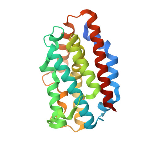
5UCA

Crystal structure of human Heme Oxygenase-2 in complex with Laurate


Experimental Data Snapshot

  • Method: X-RAY DIFFRACTION
  • Resolution: 2.12 Å
  • R-Value Free: 0.249 
  • R-Value Work: 0.193 
  • R-Value Observed: 0.195 

Starting Model: experimental
View more details

wwPDB Validation   3D Report Full Report


Ligand Structure Quality Assessment 


This is version 1.3 of the entry. See complete history


Literature

Heme Oxygenase 2 Binds Myristate to Regulate Retrovirus Assembly and TLR4 Signaling.

Zhu, Y.Luo, S.Sabo, Y.Wang, C.Tong, L.Goff, S.P.

(2017) Cell Host Microbe 21: 220-230

  • DOI: https://doi.org/10.1016/j.chom.2017.01.002
  • Primary Citation of Related Structures:  
    5UC8, 5UC9, 5UCA

  • PubMed Abstract: 

    N-myristoylation is the covalent attachment of myristic acid to the N terminus of proteins in eukaryotic cells. The matrix domain (MA) of HIV-1 Gag protein is N-myristoylated and plays an important role in virus budding. In screening for host factors that interact with HIV-1 MA, we found that heme oxygenase (HO-2) specifically binds the myristate moiety of Gag. HO-2 was also found to bind TRAM, an adaptor protein for Toll-like receptor 4 (TLR4), and thereby impact both virus replication and cellular inflammatory responses. A crystal structure revealed that HO-2 binds myristate via a hydrophobic channel adjacent to the heme-binding pocket. Inhibiting HO-2 expression, or blocking myristate binding with a heme analog, led to marked increases in virus production. HO-2 deficiency caused hyperresponsive TRAM-dependent TLR4 signaling and hypersensitivity to the TLR4 ligand lipopolysaccharide. Thus, HO-2 is a cellular myristate-binding protein that negatively regulates both virus replication and host inflammatory responses.


  • Organizational Affiliation

    Howard Hughes Medical Institute, Department of Biochemistry and Molecular Biophysics, and Department of Microbiology and Immunology, Columbia University, New York, NY 10032, USA.


Macromolecules
Find similar proteins by:  (by identity cutoff)  |  3D Structure
Entity ID: 1
MoleculeChains Sequence LengthOrganismDetailsImage
Heme oxygenase 2
A, B, C, D
226Homo sapiensMutation(s): 0 
Gene Names: HMOX2HO2
EC: 1.14.14.18
UniProt & NIH Common Fund Data Resources
Find proteins for P30519 (Homo sapiens)
Explore P30519 
Go to UniProtKB:  P30519
PHAROS:  P30519
GTEx:  ENSG00000103415 
Entity Groups  
Sequence Clusters30% Identity50% Identity70% Identity90% Identity95% Identity100% Identity
UniProt GroupP30519
Sequence Annotations
Expand
  • Reference Sequence
Experimental Data & Validation

Experimental Data

  • Method: X-RAY DIFFRACTION
  • Resolution: 2.12 Å
  • R-Value Free: 0.249 
  • R-Value Work: 0.193 
  • R-Value Observed: 0.195 
  • Space Group: P 21 21 21
Unit Cell:
Length ( Å )Angle ( ˚ )
a = 77.363α = 90
b = 83.92β = 90
c = 138.297γ = 90
Software Package:
Software NamePurpose
HKL-2000data scaling
PHENIXrefinement
PDB_EXTRACTdata extraction
HKL-2000data reduction
PHENIXphasing
DENZOdata reduction
SCALEPACKdata scaling

Structure Validation

View Full Validation Report


Ligand Structure Quality Assessment 


Entry History 

Deposition Data

Revision History  (Full details and data files)

  • Version 1.0: 2017-02-15
    Type: Initial release
  • Version 1.1: 2017-02-22
    Changes: Database references
  • Version 1.2: 2017-11-22
    Changes: Refinement description
  • Version 1.3: 2023-10-04
    Changes: Data collection, Database references, Refinement description