5UCA | pdb_00005uca

Crystal structure of human Heme Oxygenase-2 in complex with Laurate


Experimental Data Snapshot

  • Method: X-RAY DIFFRACTION
  • Resolution: 2.12 Å
  • R-Value Free: 
    0.249 (Depositor), 0.249 (DCC) 
  • R-Value Work: 
    0.193 (Depositor), 0.194 (DCC) 
  • R-Value Observed: 
    0.195 (Depositor) 

Starting Model: experimental
View more details

wwPDB Validation 3D Report Full Report

Validation slider image for 5UCA

Ligand Structure Quality Assessment 


This is version 1.3 of the entry. See complete history

Literature

Heme Oxygenase 2 Binds Myristate to Regulate Retrovirus Assembly and TLR4 Signaling.

Zhu, Y.Luo, S.Sabo, Y.Wang, C.Tong, L.Goff, S.P.

(2017) Cell Host Microbe 21: 220-230

  • DOI: https://doi.org/10.1016/j.chom.2017.01.002
  • Primary Citation Related Structures: 
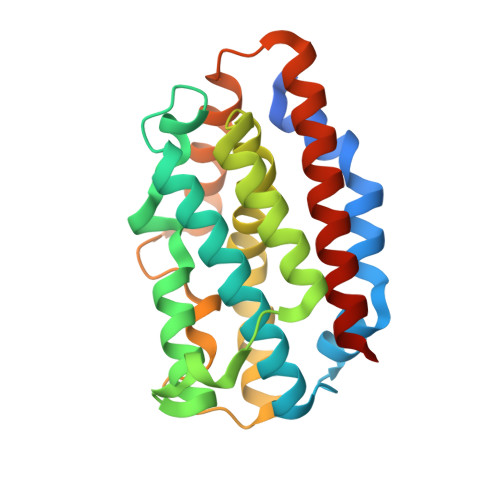
    5UC8, 5UC9, 5UCA

  • PubMed Abstract: 

    N-myristoylation is the covalent attachment of myristic acid to the N terminus of proteins in eukaryotic cells. The matrix domain (MA) of HIV-1 Gag protein is N-myristoylated and plays an important role in virus budding. In screening for host factors that interact with HIV-1 MA, we found that heme oxygenase (HO-2) specifically binds the myristate moiety of Gag. HO-2 was also found to bind TRAM, an adaptor protein for Toll-like receptor 4 (TLR4), and thereby impact both virus replication and cellular inflammatory responses. A crystal structure revealed that HO-2 binds myristate via a hydrophobic channel adjacent to the heme-binding pocket. Inhibiting HO-2 expression, or blocking myristate binding with a heme analog, led to marked increases in virus production. HO-2 deficiency caused hyperresponsive TRAM-dependent TLR4 signaling and hypersensitivity to the TLR4 ligand lipopolysaccharide. Thus, HO-2 is a cellular myristate-binding protein that negatively regulates both virus replication and host inflammatory responses.


  • Organizational Affiliation
    • Howard Hughes Medical Institute, Department of Biochemistry and Molecular Biophysics, and Department of Microbiology and Immunology, Columbia University, New York, NY 10032, USA.

Macromolecule Content 

  • Total Structure Weight: 105.75 kDa 
  • Atom Count: 7,371 
  • Modeled Residue Count: 839 
  • Deposited Residue Count: 904 
  • Unique protein chains: 1

Macromolecules

Find similar proteins by:|  3D Structure
Entity ID: 1
MoleculeChains  Sequence LengthOrganismDetailsImage
Heme oxygenase 2
A, B, C, D
226Homo sapiensMutation(s): 0 
Gene Names: HMOX2HO2
EC: 1.14.14.18
UniProt & NIH Common Fund Data Resources
Find proteins for P30519 (Homo sapiens)
Explore P30519 
Go to UniProtKB:  P30519
PHAROS:  P30519
GTEx:  ENSG00000103415 
Entity Groups
Sequence Clusters30% Identity50% Identity70% Identity90% Identity95% Identity100% Identity
UniProt GroupP30519
Sequence Annotations
Expand
Reference Sequence

Experimental Data & Validation

Experimental Data

  • Method: X-RAY DIFFRACTION
  • Resolution: 2.12 Å
  • R-Value Free:  0.249 (Depositor), 0.249 (DCC) 
  • R-Value Work:  0.193 (Depositor), 0.194 (DCC) 
  • R-Value Observed: 0.195 (Depositor) 
Space Group: P 21 21 21
Unit Cell:
Length ( Å )Angle ( ˚ )
a = 77.363α = 90
b = 83.92β = 90
c = 138.297γ = 90
Software Package:
Software NamePurpose
HKL-2000data scaling
PHENIXrefinement
PDB_EXTRACTdata extraction
HKL-2000data reduction
PHENIXphasing
DENZOdata reduction
SCALEPACKdata scaling

Structure Validation

View Full Validation Report


Ligand Structure Quality Assessment 


Entry History 

Deposition Data

Revision History  (Full details and data files)

  • Version 1.0: 2017-02-15
    Type: Initial release
  • Version 1.1: 2017-02-22
    Changes: Database references
  • Version 1.2: 2017-11-22
    Changes: Refinement description
  • Version 1.3: 2023-10-04
    Changes: Data collection, Database references, Refinement description