5U35 | pdb_00005u35

Crystal structure of a de novo designed protein with curved beta-sheet


Experimental Data Snapshot

  • Method: X-RAY DIFFRACTION
  • Resolution: 1.80 Å
  • R-Value Free: 
    0.247 (Depositor), 0.247 (DCC) 
  • R-Value Work: 
    0.201 (Depositor), 0.202 (DCC) 
  • R-Value Observed: 
    0.206 (Depositor) 

wwPDB Validation 3D Report Full Report

Validation slider image for 5U35

This is version 1.2 of the entry. See complete history

Literature

Principles for designing proteins with cavities formed by curved beta sheets.

Marcos, E.Basanta, B.Chidyausiku, T.M.Tang, Y.Oberdorfer, G.Liu, G.Swapna, G.V.Guan, R.Silva, D.A.Dou, J.Pereira, J.H.Xiao, R.Sankaran, B.Zwart, P.H.Montelione, G.T.Baker, D.

(2017) Science 355: 201-206

  • DOI: https://doi.org/10.1126/science.aah7389
  • Primary Citation Related Structures: 
    5KPE, 5KPH, 5L33, 5TPH, 5TPJ, 5TRV, 5TS4, 5U35

  • PubMed Abstract: 

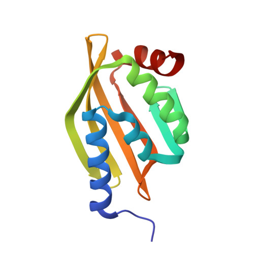
    Active sites and ligand-binding cavities in native proteins are often formed by curved β sheets, and the ability to control β-sheet curvature would allow design of binding proteins with cavities customized to specific ligands. Toward this end, we investigated the mechanisms controlling β-sheet curvature by studying the geometry of β sheets in naturally occurring protein structures and folding simulations. The principles emerging from this analysis were used to design, de novo, a series of proteins with curved β sheets topped with α helices. Nuclear magnetic resonance and crystal structures of the designs closely match the computational models, showing that β-sheet curvature can be controlled with atomic-level accuracy. Our approach enables the design of proteins with cavities and provides a route to custom design ligand-binding and catalytic sites.


  • Organizational Affiliation
    • Department of Biochemistry, University of Washington, Seattle, WA 98195, USA.

Macromolecule Content 

  • Total Structure Weight: 29.67 kDa 
  • Atom Count: 2,125 
  • Modeled Residue Count: 246 
  • Deposited Residue Count: 250 
  • Unique protein chains: 1

Macromolecules

Find similar proteins by:|  3D Structure
Entity ID: 1
MoleculeChains  Sequence LengthOrganismDetailsImage
De novo NTF2 with large cavity
A, B
125synthetic constructMutation(s): 0 

Small Molecules

Ligands 5 Unique
IDChains Name / Formula / InChI Key2D Diagram3D Interactions
PG6

Query on PG6


Download:Ideal Coordinates CCD File
I [auth B]1-(2-METHOXY-ETHOXY)-2-{2-[2-(2-METHOXY-ETHOXY]-ETHOXY}-ETHANE
C12 H26 O6
DMDPGPKXQDIQQG-UHFFFAOYSA-N
PG4

Query on PG4


Download:Ideal Coordinates CCD File
H [auth A]TETRAETHYLENE GLYCOL
C8 H18 O5
UWHCKJMYHZGTIT-UHFFFAOYSA-N
MXE

Query on MXE


Download:Ideal Coordinates CCD File
C [auth A],
D [auth A],
E [auth A],
F [auth A],
J [auth B]
2-METHOXYETHANOL
C3 H8 O2
XNWFRZJHXBZDAG-UHFFFAOYSA-N
CL

Query on CL


Download:Ideal Coordinates CCD File
G [auth A],
K [auth B]
CHLORIDE ION
Cl
VEXZGXHMUGYJMC-UHFFFAOYSA-M
NA

Query on NA


Download:Ideal Coordinates CCD File
L [auth B]SODIUM ION
Na
FKNQFGJONOIPTF-UHFFFAOYSA-N

Experimental Data & Validation

Experimental Data

  • Method: X-RAY DIFFRACTION
  • Resolution: 1.80 Å
  • R-Value Free:  0.247 (Depositor), 0.247 (DCC) 
  • R-Value Work:  0.201 (Depositor), 0.202 (DCC) 
  • R-Value Observed: 0.206 (Depositor) 
Space Group: P 1 21 1
Unit Cell:
Length ( Å )Angle ( ˚ )
a = 38.01α = 90
b = 33.2β = 91.78
c = 86.59γ = 90
Software Package:
Software NamePurpose
PHENIXrefinement
iMOSFLMdata reduction
SCALAdata scaling
PHASERphasing

Structure Validation

View Full Validation Report


Entry History 

Deposition Data

Revision History  (Full details and data files)

  • Version 1.0: 2017-01-25
    Type: Initial release
  • Version 1.1: 2017-02-01
    Changes: Database references
  • Version 1.2: 2024-03-06
    Changes: Data collection, Database references, Derived calculations