5TL5 | pdb_00005tl5

COMPLEX BETWEEN HUMAN CD27 AND ANTIBODY M2177


Experimental Data Snapshot

  • Method: X-RAY DIFFRACTION
  • Resolution: 1.80 Å
  • R-Value Free: 
    0.232 (Depositor), 0.233 (DCC) 
  • R-Value Work: 
    0.197 (Depositor), 0.199 (DCC) 
  • R-Value Observed: 
    0.197 (Depositor) 

Starting Model: experimental
View more details

wwPDB Validation 3D Report Full Report

Validation slider image for 5TL5

Ligand Structure Quality Assessment 


This is version 2.2 of the entry. See complete history

Literature

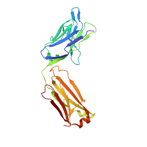
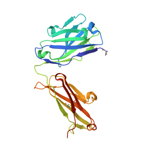
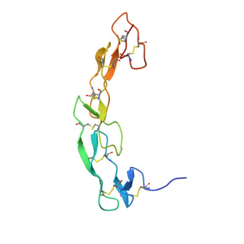
Crystal structure of CD27 in complex with a neutralizing noncompeting antibody.

Teplyakov, A.Obmolova, G.Malia, T.J.Gilliland, G.L.

(2017) Acta Crystallogr F Struct Biol Commun 73: 294-299

  • DOI: https://doi.org/10.1107/S2053230X17005957
  • Primary Citation Related Structures: 
    5TL5

  • PubMed Abstract: 

    CD27 is a T-cell and B-cell co-stimulatory glycoprotein of the tumor necrosis factor (TNF) receptor superfamily that is dependent on the availability of the TNF-like ligand CD70. Therapeutic approaches to treating autoimmune diseases and cancers with antagonistic and agonistic anti-CD27 monoclonal antibodies (mAbs), respectively, have recently been developed. Mouse anti-human CD27 mAb 2177 shows potency in neutralizing CD70-induced signaling; however, it does not block the binding of soluble CD70. To provide insight into the mechanism of action of the mAb, the crystal structure of the CD27 extracellular domain in complex with the Fab fragment of mAb 2177 was determined at 1.8 Å resolution. CD27 exhibits the assembly of cysteine-rich domains characteristic of the TNF receptor superfamily. The structure reveals a unique binding site of mAb 2177 at the edge of the receptor molecule, which allows the mAb to sterically block the cell-bound form of CD70 from reaching CD27 while leaving the ligand epitope clear. This mode of action suggests a potential dual use of mAb 2177 either as an antagonist or as an agonist.


  • Organizational Affiliation
    • Janssen Research and Development LLC, 1400 McKean Road, Spring House, PA 19477, USA.

Macromolecule Content 

  • Total Structure Weight: 60.76 kDa 
  • Atom Count: 4,402 
  • Modeled Residue Count: 538 
  • Deposited Residue Count: 556 
  • Unique protein chains: 3

Macromolecules

Find similar proteins by:|  3D Structure
Entity ID: 1
MoleculeChains  Sequence LengthOrganismDetailsImage
M2177 LIGHT CHAINA [auth L]218Mus musculusHomo sapiens
This entity is chimeric
Mutation(s): 0 
Find similar proteins by:|  3D Structure
Entity ID: 2
MoleculeChains  Sequence LengthOrganismDetailsImage
M2177 HEAVY CHAINB [auth H]229Mus musculusHomo sapiens
This entity is chimeric
Mutation(s): 0 
Find similar proteins by:|  3D Structure
Entity ID: 3
MoleculeChains  Sequence LengthOrganismDetailsImage
CD27 antigenC [auth A]109Homo sapiensMutation(s): 0 
Gene Names: CD27TNFRSF7
UniProt & NIH Common Fund Data Resources
Find proteins for P26842 (Homo sapiens)
Explore P26842 
Go to UniProtKB:  P26842
PHAROS:  P26842
GTEx:  ENSG00000139193 
Entity Groups
Sequence Clusters30% Identity50% Identity70% Identity90% Identity95% Identity100% Identity
UniProt GroupP26842
Glycosylation
Glycosylation Sites: 1Go to GlyGen: P26842-1
Sequence Annotations
Expand
Reference Sequence

Small Molecules

Modified Residues  1 Unique
IDChains TypeFormula2D DiagramParent
PCA
Query on PCA
B [auth H]L-PEPTIDE LINKINGC5 H7 N O3GLN

Experimental Data & Validation

Experimental Data

  • Method: X-RAY DIFFRACTION
  • Resolution: 1.80 Å
  • R-Value Free:  0.232 (Depositor), 0.233 (DCC) 
  • R-Value Work:  0.197 (Depositor), 0.199 (DCC) 
  • R-Value Observed: 0.197 (Depositor) 
Space Group: C 1 2 1
Unit Cell:
Length ( Å )Angle ( ˚ )
a = 274.2α = 90
b = 37.41β = 98.82
c = 61.08γ = 90
Software Package:
Software NamePurpose
XDSdata reduction
XDSdata scaling
REFMACrefinement
PHASERphasing

Structure Validation

View Full Validation Report


Ligand Structure Quality Assessment 


Entry History 

Deposition Data

Revision History  (Full details and data files)

  • Version 1.0: 2017-06-14
    Type: Initial release
  • Version 2.0: 2020-07-29
    Type: Remediation
    Reason: Carbohydrate remediation
    Changes: Data collection, Derived calculations, Polymer sequence, Structure summary
  • Version 2.1: 2023-10-04
    Changes: Data collection, Database references, Refinement description, Structure summary
  • Version 2.2: 2024-10-23
    Changes: Structure summary