5NV3 | pdb_00005nv3

Structure of Rubisco from Rhodobacter sphaeroides in complex with CABP


Experimental Data Snapshot

  • Method: ELECTRON MICROSCOPY
  • Resolution: 3.39 Å
  • Aggregation State: PARTICLE 
  • Reconstruction Method: SINGLE PARTICLE 

wwPDB Validation 3D Report Full Report

Validation slider image for 5NV3

This is version 1.3 of the entry. See complete history

Literature

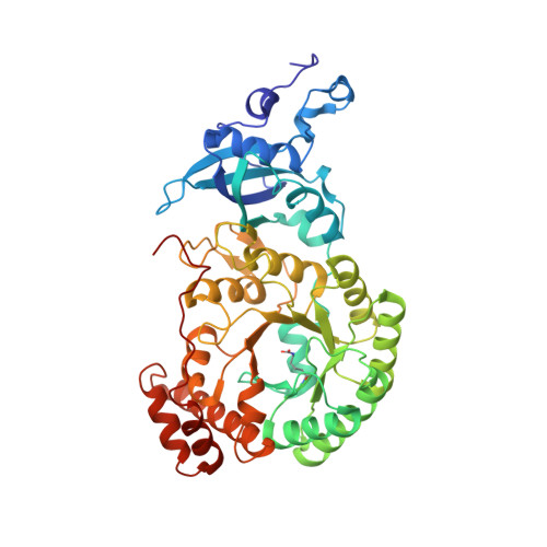
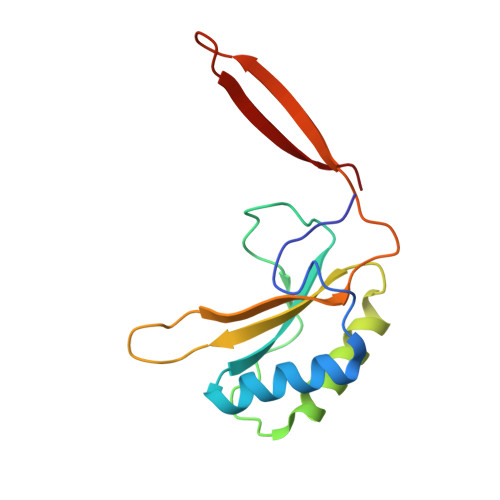
Mechanism of Enzyme Repair by the AAA(+) Chaperone Rubisco Activase.

Bhat, J.Y.Milicic, G.Thieulin-Pardo, G.Bracher, A.Maxwell, A.Ciniawsky, S.Mueller-Cajar, O.Engen, J.R.Hartl, F.U.Wendler, P.Hayer-Hartl, M.

(2017) Mol Cell 67: 744-756.e6

  • DOI: https://doi.org/10.1016/j.molcel.2017.07.004
  • Primary Citation Related Structures: 
    5NV3

  • PubMed Abstract: 

    How AAA+ chaperones conformationally remodel specific target proteins in an ATP-dependent manner is not well understood. Here, we investigated the mechanism of the AAA+ protein Rubisco activase (Rca) in metabolic repair of the photosynthetic enzyme Rubisco, a complex of eight large (RbcL) and eight small (RbcS) subunits containing eight catalytic sites. Rubisco is prone to inhibition by tight-binding sugar phosphates, whose removal is catalyzed by Rca. We engineered a stable Rca hexamer ring and analyzed its functional interaction with Rubisco. Hydrogen/deuterium exchange and chemical crosslinking showed that Rca structurally destabilizes elements of the Rubisco active site with remarkable selectivity. Cryo-electron microscopy revealed that Rca docks onto Rubisco over one active site at a time, positioning the C-terminal strand of RbcL, which stabilizes the catalytic center, for access to the Rca hexamer pore. The pulling force of Rca is fine-tuned to avoid global destabilization and allow for precise enzyme repair.


  • Organizational Affiliation
    • Department of Cellular Biochemistry, Max Planck Institute of Biochemistry, Am Klopferspitz 18, 82152 Martinsried, Germany.

Macromolecule Content 

  • Total Structure Weight: 538.66 kDa 
  • Atom Count: 37,816 
  • Modeled Residue Count: 4,768 
  • Deposited Residue Count: 4,768 
  • Unique protein chains: 2

Macromolecules

Find similar proteins by:|  3D Structure
Entity ID: 1
MoleculeChains  Sequence LengthOrganismDetailsImage
Ribulose bisphosphate carboxylase large chain467Cereibacter sphaeroidesMutation(s): 0 
Gene Names: cbbLcbbL1rbcL
EC: 4.1.1.39
UniProt
Find proteins for P27997 (Cereibacter sphaeroides)
Explore P27997 
Go to UniProtKB:  P27997
Entity Groups
Sequence Clusters30% Identity50% Identity70% Identity90% Identity95% Identity100% Identity
UniProt GroupP27997
Sequence Annotations
Expand
Reference Sequence
Find similar proteins by:|  3D Structure
Entity ID: 2
MoleculeChains  Sequence LengthOrganismDetailsImage
Ribulose bisphosphate carboxylase small chain 1129Cereibacter sphaeroidesMutation(s): 0 
Gene Names: cbbSrbcS
EC: 4.1.1.39
UniProt
Find proteins for P27998 (Cereibacter sphaeroides)
Explore P27998 
Go to UniProtKB:  P27998
Entity Groups
Sequence Clusters30% Identity50% Identity70% Identity90% Identity95% Identity100% Identity
UniProt GroupP27998
Sequence Annotations
Expand
Reference Sequence

Small Molecules

Ligands 2 Unique
IDChains Name / Formula / InChI Key2D Diagram3D Interactions
CAP

Query on CAP


Download:Ideal Coordinates CCD File
AA [auth F]
CA [auth G]
EA [auth H]
Q [auth A]
S [auth B]
AA [auth F],
CA [auth G],
EA [auth H],
Q [auth A],
S [auth B],
U [auth C],
W [auth D],
Y [auth E]
2-CARBOXYARABINITOL-1,5-DIPHOSPHATE
C6 H14 O13 P2
ITHCSGCUQDMYAI-ZMIZWQJLSA-N
MG

Query on MG


Download:Ideal Coordinates CCD File
BA [auth F]
DA [auth G]
FA [auth H]
R [auth A]
T [auth B]
BA [auth F],
DA [auth G],
FA [auth H],
R [auth A],
T [auth B],
V [auth C],
X [auth D],
Z [auth E]
MAGNESIUM ION
Mg
JLVVSXFLKOJNIY-UHFFFAOYSA-N
Modified Residues  1 Unique
IDChains TypeFormula2D DiagramParent
KCX
Query on KCX
A
C [auth B]
E [auth C]
G [auth D]
I [auth E]
A,
C [auth B],
E [auth C],
G [auth D],
I [auth E],
K [auth F],
M [auth G],
O [auth H]
L-PEPTIDE LINKINGC7 H14 N2 O4LYS

Experimental Data & Validation

Experimental Data

  • Method: ELECTRON MICROSCOPY
  • Resolution: 3.39 Å
  • Aggregation State: PARTICLE 
  • Reconstruction Method: SINGLE PARTICLE 
EM Software:
TaskSoftware PackageVersion
MODEL REFINEMENTREFMAC5.8.0155
RECONSTRUCTIONRELION

Structure Validation

View Full Validation Report


Entry History 

Deposition Data

Revision History  (Full details and data files)

  • Version 1.0: 2017-07-26
    Type: Initial release
  • Version 1.1: 2017-08-23
    Changes: Database references, Refinement description
  • Version 1.2: 2017-09-20
    Changes: Database references
  • Version 1.3: 2025-04-09
    Changes: Data collection, Database references, Derived calculations, Structure summary