5N7O

EthR2 in complex with SMARt-420 compound


Experimental Data Snapshot

  • Method: X-RAY DIFFRACTION
  • Resolution: 2.30 Å
  • R-Value Free: 0.248 
  • R-Value Work: 0.215 
  • R-Value Observed: 0.217 

Starting Model: experimental
View more details

wwPDB Validation   3D Report Full Report


Ligand Structure Quality Assessment 


This is version 1.2 of the entry. See complete history


Literature

Structural analysis of the interaction between spiroisoxazoline SMARt-420 and the Mycobacterium tuberculosis repressor EthR2.

Wohlkonig, A.Remaut, H.Moune, M.Tanina, A.Meyer, F.Desroses, M.Steyaert, J.Willand, N.Baulard, A.R.Wintjens, R.

(2017) Biochem Biophys Res Commun 487: 403-408

  • DOI: https://doi.org/10.1016/j.bbrc.2017.04.074
  • Primary Citation of Related Structures:  
    5N7O

  • PubMed Abstract: 

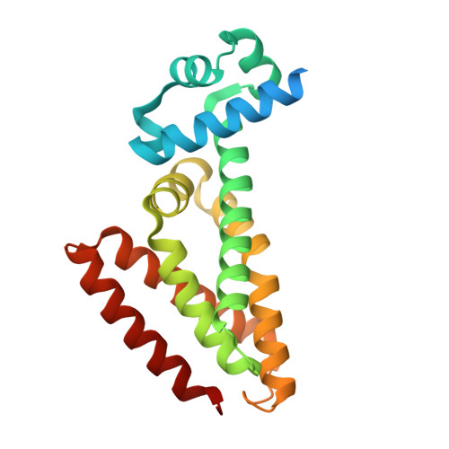
    Inhibition of transcriptional regulators of bacterial pathogens with the aim of reprogramming their metabolism to modify their antibiotic susceptibility constitutes a promising therapeutic strategy. One example is the bio-activation of the anti-tubercular pro-drug ethionamide, which activity could be enhanced by inhibiting the transcriptional repressor EthR. Recently, we discovered that inhibition of a second transcriptional repressor, EthR2, leads to the awakening of a new ethionamide bio-activation pathway. The x-ray structure of EthR2 was solved at 2.3 Å resolution in complex with a compound called SMARt-420 (Small Molecule Aborting Resistance). Detailed comparison and structural analysis revealed interesting insights for the upcoming structure-based design of EthR2 inhibitors as an alternative to revert ethionamide resistance in Mycobacterium tuberculosis.


  • Organizational Affiliation

    Center for Structural Biology, Vlaams Instituut voor Biotechnology (VIB), Pleinlaan 2, B-1050 Brussels, Belgium; Structural Biology Brussels, Vrije Universiteit Brussel (VUB), Pleinlaan 2, B-1050 Brussels, Belgium.


Macromolecules
Find similar proteins by:  (by identity cutoff)  |  3D Structure
Entity ID: 1
MoleculeChains Sequence LengthOrganismDetailsImage
Probable transcriptional regulatory protein
A, B
221Mycobacterium tuberculosis H37RvMutation(s): 0 
Gene Names: Rv0078LH57_00450
UniProt
Find proteins for O53623 (Mycobacterium tuberculosis (strain ATCC 25618 / H37Rv))
Explore O53623 
Go to UniProtKB:  O53623
Entity Groups  
Sequence Clusters30% Identity50% Identity70% Identity90% Identity95% Identity100% Identity
UniProt GroupO53623
Sequence Annotations
Expand
  • Reference Sequence
Small Molecules
Ligands 1 Unique
IDChains Name / Formula / InChI Key2D Diagram3D Interactions
69Y
Query on 69Y

Download Ideal Coordinates CCD File 
C [auth A],
D [auth B]
4,4,4-trifluoro-1-(3-phenyl-1-oxa-2,8-diazaspiro[4.5]dec-2-en-8-yl)butan-1-one
C17 H19 F3 N2 O2
IBVDSAFESRHIBV-UHFFFAOYSA-N
Experimental Data & Validation

Experimental Data

  • Method: X-RAY DIFFRACTION
  • Resolution: 2.30 Å
  • R-Value Free: 0.248 
  • R-Value Work: 0.215 
  • R-Value Observed: 0.217 
  • Space Group: P 21 21 21
Unit Cell:
Length ( Å )Angle ( ˚ )
a = 59.4α = 90
b = 74.29β = 90
c = 91.5γ = 90
Software Package:
Software NamePurpose
REFMACrefinement
XDSdata reduction
XDSdata scaling
MOLREPphasing

Structure Validation

View Full Validation Report


Ligand Structure Quality Assessment 


Entry History 

Deposition Data

Revision History  (Full details and data files)

  • Version 1.0: 2017-04-26
    Type: Initial release
  • Version 1.1: 2017-05-10
    Changes: Database references
  • Version 1.2: 2024-01-17
    Changes: Data collection, Database references, Refinement description