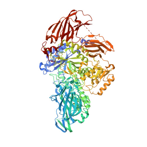
5MQM

Glycoside hydrolase BT_0986


Experimental Data Snapshot

  • Method: X-RAY DIFFRACTION
  • Resolution: 2.11 Å
  • R-Value Free: 0.240 
  • R-Value Work: 0.199 
  • R-Value Observed: 0.201 

wwPDB Validation   3D Report Full Report


Ligand Structure Quality Assessment 


This is version 1.4 of the entry. See complete history


Literature

Complex pectin metabolism by gut bacteria reveals novel catalytic functions.

Ndeh, D.Rogowski, A.Cartmell, A.Luis, A.S.Basle, A.Gray, J.Venditto, I.Briggs, J.Zhang, X.Labourel, A.Terrapon, N.Buffetto, F.Nepogodiev, S.Xiao, Y.Field, R.A.Zhu, Y.O'Neill, M.A.Urbanowicz, B.R.York, W.S.Davies, G.J.Abbott, D.W.Ralet, M.C.Martens, E.C.Henrissat, B.Gilbert, H.J.

(2017) Nature 544: 65-70

  • DOI: https://doi.org/10.1038/nature21725
  • Primary Citation of Related Structures:  
    5MQM, 5MQN, 5MQO, 5MQR, 5MQS, 5MSX, 5MSY, 5MT2, 5MUI, 5MUJ, 5MWK

  • PubMed Abstract: 

    The metabolism of carbohydrate polymers drives microbial diversity in the human gut microbiota. It is unclear, however, whether bacterial consortia or single organisms are required to depolymerize highly complex glycans. Here we show that the gut bacterium Bacteroides thetaiotaomicron uses the most structurally complex glycan known: the plant pectic polysaccharide rhamnogalacturonan-II, cleaving all but 1 of its 21 distinct glycosidic linkages. The deconstruction of rhamnogalacturonan-II side chains and backbone are coordinated to overcome steric constraints, and the degradation involves previously undiscovered enzyme families and catalytic activities. The degradation system informs revision of the current structural model of rhamnogalacturonan-II and highlights how individual gut bacteria orchestrate manifold enzymes to metabolize the most challenging glycan in the human diet.


  • Organizational Affiliation

    Institute for Cell and Molecular Biosciences, Newcastle University, Newcastle upon Tyne NE2 4HH, U.K.


Macromolecules
Find similar proteins by:  (by identity cutoff)  |  3D Structure
Entity ID: 1
MoleculeChains Sequence LengthOrganismDetailsImage
Glycosyl hydrolases family 2, sugar binding domain1,105Bacteroides thetaiotaomicronMutation(s): 0 
Gene Names: Btheta7330_02599
UniProt
Find proteins for Q8A931 (Bacteroides thetaiotaomicron (strain ATCC 29148 / DSM 2079 / JCM 5827 / CCUG 10774 / NCTC 10582 / VPI-5482 / E50))
Explore Q8A931 
Go to UniProtKB:  Q8A931
Entity Groups  
Sequence Clusters30% Identity50% Identity70% Identity90% Identity95% Identity100% Identity
UniProt GroupQ8A931
Sequence Annotations
Expand
  • Reference Sequence
Small Molecules
Ligands 4 Unique
IDChains Name / Formula / InChI Key2D Diagram3D Interactions
PG4
Query on PG4

Download Ideal Coordinates CCD File 
F [auth A],
G [auth A]
TETRAETHYLENE GLYCOL
C8 H18 O5
UWHCKJMYHZGTIT-UHFFFAOYSA-N
RPQ
Query on RPQ

Download Ideal Coordinates CCD File 
E [auth A]D-rhamnopyranose tetrazole
C6 H10 N4 O3
RGTARVZVTUMUSY-QMKXCQHVSA-N
PGE
Query on PGE

Download Ideal Coordinates CCD File 
H [auth A],
I [auth A]
TRIETHYLENE GLYCOL
C6 H14 O4
ZIBGPFATKBEMQZ-UHFFFAOYSA-N
CA
Query on CA

Download Ideal Coordinates CCD File 
B [auth A],
C [auth A],
D [auth A]
CALCIUM ION
Ca
BHPQYMZQTOCNFJ-UHFFFAOYSA-N
Experimental Data & Validation

Experimental Data

  • Method: X-RAY DIFFRACTION
  • Resolution: 2.11 Å
  • R-Value Free: 0.240 
  • R-Value Work: 0.199 
  • R-Value Observed: 0.201 
  • Space Group: P 1 21 1
Unit Cell:
Length ( Å )Angle ( ˚ )
a = 64.05α = 90
b = 84.81β = 99.2
c = 120.63γ = 90
Software Package:
Software NamePurpose
xia2data reduction
Aimlessdata scaling
MOLREPphasing
Cootmodel building
REFMACrefinement

Structure Validation

View Full Validation Report


Ligand Structure Quality Assessment 


Entry History & Funding Information

Deposition Data


Funding OrganizationLocationGrant Number
iotechnology and Biological Research CouncilUnited KingdomBB/K020358/1
iotechnology and Biological Research CouncilUnited KingdomBB/K001949/1
Wellcome TrustUnited KingdomWT097907MA
European Research CouncilUnited Kingdom322820

Revision History  (Full details and data files)

  • Version 1.0: 2017-03-22
    Type: Initial release
  • Version 1.1: 2017-04-05
    Changes: Database references
  • Version 1.2: 2017-04-12
    Changes: Database references
  • Version 1.3: 2017-08-23
    Changes: Author supporting evidence
  • Version 1.4: 2024-05-01
    Changes: Data collection, Database references, Refinement description