5MMX | pdb_00005mmx

ABA RECEPTOR FROM CITRUS, CSPYL1


Experimental Data Snapshot

  • Method: X-RAY DIFFRACTION
  • Resolution: 2.88 Å
  • R-Value Free: 
    0.293 (Depositor), 0.293 (DCC) 
  • R-Value Work: 
    0.226 (Depositor), 0.232 (DCC) 
  • R-Value Observed: 
    0.230 (Depositor) 

Starting Model: experimental
View more details

wwPDB Validation 3D Report Full Report

Validation slider image for 5MMX

Ligand Structure Quality Assessment 


This is version 1.4 of the entry. See complete history

Literature

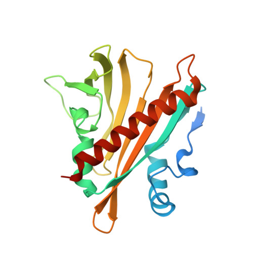
Structure of Ligand-Bound Intermediates of Crop ABA Receptors Highlights PP2C as Necessary ABA Co-receptor.

Moreno-Alvero, M.Yunta, C.Gonzalez-Guzman, M.Lozano-Juste, J.Benavente, J.L.Arbona, V.Menendez, M.Martinez-Ripoll, M.Infantes, L.Gomez-Cadenas, A.Rodriguez, P.L.Albert, A.

(2017) Mol Plant 10: 1250-1253

  • DOI: https://doi.org/10.1016/j.molp.2017.07.004
  • Primary Citation Related Structures: 
    5MMQ, 5MMX, 5MN0, 5MOA, 5MOB

  • Organizational Affiliation
    • Instituto de Química Física Rocasolano, Consejo Superior de Investigaciones Científicas, 28006 Madrid, Spain.

Macromolecule Content 

  • Total Structure Weight: 47.12 kDa 
  • Atom Count: 2,968 
  • Modeled Residue Count: 372 
  • Deposited Residue Count: 418 
  • Unique protein chains: 1

Macromolecules

Find similar proteins by:|  3D Structure
Entity ID: 1
MoleculeChains  Sequence LengthOrganismDetailsImage
CSPYL1_ABAA [auth B],
B [auth A]
209Citrus sinensisMutation(s): 0 
Gene Names: CISIN_1g046151mg
UniProt
Find proteins for A0A067E666 (Citrus sinensis)
Explore A0A067E666 
Go to UniProtKB:  A0A067E666
Entity Groups
Sequence Clusters30% Identity50% Identity70% Identity90% Identity95% Identity100% Identity
UniProt GroupA0A067E666
Sequence Annotations
Expand
Reference Sequence

Small Molecules

Ligands 1 Unique
IDChains Name / Formula / InChI Key2D Diagram3D Interactions
A8S

Query on A8S


Download:Ideal Coordinates CCD File
C [auth B],
D [auth A]
(2Z,4E)-5-[(1S)-1-hydroxy-2,6,6-trimethyl-4-oxocyclohex-2-en-1-yl]-3-methylpenta-2,4-dienoic acid
C15 H20 O4
JLIDBLDQVAYHNE-YKALOCIXSA-N

Experimental Data & Validation

Experimental Data

  • Method: X-RAY DIFFRACTION
  • Resolution: 2.88 Å
  • R-Value Free:  0.293 (Depositor), 0.293 (DCC) 
  • R-Value Work:  0.226 (Depositor), 0.232 (DCC) 
  • R-Value Observed: 0.230 (Depositor) 
Space Group: P 4 21 2
Unit Cell:
Length ( Å )Angle ( ˚ )
a = 82.613α = 90
b = 82.613β = 90
c = 126.674γ = 90
Software Package:
Software NamePurpose
PHENIXrefinement
Aimlessdata scaling
SCALAdata scaling
MOLREPphasing

Structure Validation

View Full Validation Report


Ligand Structure Quality Assessment 


Entry History 

Revision History  (Full details and data files)

  • Version 1.0: 2017-08-02
    Type: Initial release
  • Version 1.1: 2017-09-20
    Changes: Database references
  • Version 1.2: 2019-06-12
    Changes: Data collection, Structure summary
  • Version 1.3: 2019-08-14
    Changes: Data collection
  • Version 1.4: 2024-01-17
    Changes: Data collection, Database references, Refinement description, Structure summary