5KWF | pdb_00005kwf

Joint X-ray Neutron Structure of Cholesterol Oxidase


Experimental Data Snapshot

  • Method: X-RAY DIFFRACTION
  • Resolution: 1.50 Å
  • R-Value Free: 
    0.220 (Depositor), 0.164 (DCC) 
  • R-Value Work: 
    0.190 (Depositor), 0.138 (DCC) 
  • R-Value Observed: 
    0.192 (Depositor) 

  • Method: NEUTRON DIFFRACTION
  • Resolution: 2.21 Å
  • R-Value Free: 
    0.312 (Depositor) 
  • R-Value Work: 
    0.283 (Depositor) 
  • R-Value Observed: 
    0.285 (Depositor) 

wwPDB Validation 3D Report Full Report

Validation slider image for 5KWF

This is version 1.2 of the entry. See complete history

Literature

An extended N-H bond, driven by a conserved second-order interaction, orients the flavin N5 orbital in cholesterol oxidase.

Golden, E.Yu, L.J.Meilleur, F.Blakeley, M.P.Duff, A.P.Karton, A.Vrielink, A.

(2017) Sci Rep 7: 40517-40517

  • DOI: https://doi.org/10.1038/srep40517
  • Primary Citation Related Structures: 
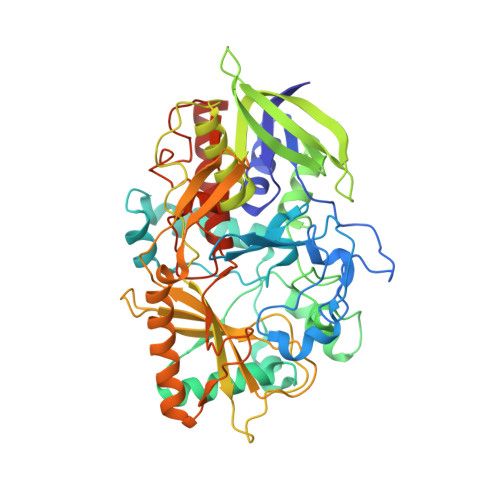
    5KWF

  • PubMed Abstract: 

    The protein microenvironment surrounding the flavin cofactor in flavoenzymes is key to the efficiency and diversity of reactions catalysed by this class of enzymes. X-ray diffraction structures of oxidoreductase flavoenzymes have revealed recurrent features which facilitate catalysis, such as a hydrogen bond between a main chain nitrogen atom and the flavin redox center (N5). A neutron diffraction study of cholesterol oxidase has revealed an unusual elongated main chain nitrogen to hydrogen bond distance positioning the hydrogen atom towards the flavin N5 reactive center. Investigation of the structural features which could cause such an unusual occurrence revealed a positively charged lysine side chain, conserved in other flavin mediated oxidoreductases, in a second shell away from the FAD cofactor acting to polarize the peptide bond through interaction with the carbonyl oxygen atom. Double-hybrid density functional theory calculations confirm that this electrostatic arrangement affects the N-H bond length in the region of the flavin reactive center. We propose a novel second-order partial-charge interaction network which enables the correct orientation of the hydride receiving orbital of N5. The implications of these observations for flavin mediated redox chemistry are discussed.


  • Organizational Affiliation
    • School of Chemistry and Biochemistry, University of Western Australia, Crawley, Western Australia, 6009 Australia.

Macromolecule Content 

  • Total Structure Weight: 56.58 kDa 
  • Atom Count: 4,145 
  • Modeled Residue Count: 499 
  • Deposited Residue Count: 510 
  • Unique protein chains: 1

Macromolecules

Find similar proteins by:|  3D Structure
Entity ID: 1
MoleculeChains  Sequence LengthOrganismDetailsImage
Cholesterol oxidase510Streptomyces sp. SA-COOMutation(s): 0 
Gene Names: choA
EC: 1.1.3.6 (PDB Primary Data), 5.3.3.1 (PDB Primary Data)
UniProt
Find proteins for P12676 (Streptomyces sp. (strain SA-COO))
Explore P12676 
Go to UniProtKB:  P12676
Entity Groups
Sequence Clusters30% Identity50% Identity70% Identity90% Identity95% Identity100% Identity
UniProt GroupP12676
Sequence Annotations
Expand
Reference Sequence

Small Molecules

Ligands 1 Unique
IDChains Name / Formula / InChI Key2D Diagram3D Interactions
FAD

Query on FAD


Download:Ideal Coordinates CCD File
B [auth A]FLAVIN-ADENINE DINUCLEOTIDE
C27 H33 N9 O15 P2
VWWQXMAJTJZDQX-UYBVJOGSSA-N

Experimental Data & Validation

Experimental Data

  • Method: X-RAY DIFFRACTION
  • Resolution: 1.50 Å
  • R-Value Free:  0.220 (Depositor), 0.164 (DCC) 
  • R-Value Work:  0.190 (Depositor), 0.138 (DCC) 
  • R-Value Observed: 0.192 (Depositor) 
  • Method: NEUTRON DIFFRACTION
  • Resolution: 2.21 Å
  • R-Value Free:  0.312 (Depositor) 
  • R-Value Work:  0.283 (Depositor) 
  • R-Value Observed: 0.285 (Depositor) 
Space Group: P 1 21 1
Unit Cell:
Length ( Å )Angle ( ˚ )
a = 51.605α = 90
b = 74.084β = 105.21
c = 63.828γ = 90
Software Package:
Software NamePurpose
PHENIXrefinement
PDB_EXTRACTdata extraction
HKL-3000data reduction
SCALAdata scaling
PHASERphasing

Structure Validation

View Full Validation Report


Entry History 

Deposition Data

Revision History  (Full details and data files)

  • Version 1.0: 2017-02-01
    Type: Initial release
  • Version 1.1: 2017-09-27
    Changes: Data collection
  • Version 1.2: 2024-03-06
    Changes: Data collection, Database references