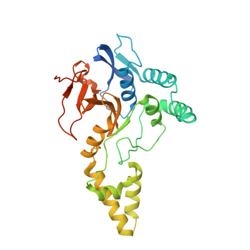
5JKN | pdb_00005jkn

Crystal structure of deubiquitinase MINDY-1


Experimental Data Snapshot

  • Method: X-RAY DIFFRACTION
  • Resolution: 3.00 Å
  • R-Value Free: 
    0.242 (Depositor), 0.247 (DCC) 
  • R-Value Work: 
    0.197 (Depositor), 0.200 (DCC) 
  • R-Value Observed: 
    0.199 (Depositor) 

Starting Model: experimental
View more details

wwPDB Validation 3D Report Full Report

Validation slider image for 5JKN

This is version 1.4 of the entry. See complete history. 

Literature

MINDY-1 Is a Member of an Evolutionarily Conserved and Structurally Distinct New Family of Deubiquitinating Enzymes.

Abdul Rehman, S.A., Kristariyanto, Y.A., Choi, S.Y., Nkosi, P.J., Weidlich, S., Labib, K., Hofmann, K., Kulathu, Y.

(2016) Mol Cell 63: 146-155

  • DOI: https://doi.org/10.1016/j.molcel.2016.05.009
  • Primary Citation Related Structures: 
    5JKN, 5JQS

  • PubMed Abstract: 

    Deubiquitinating enzymes (DUBs) remove ubiquitin (Ub) from Ub-conjugated substrates to regulate the functional outcome of ubiquitylation. Here we report the discovery of a new family of DUBs, which we have named MINDY (motif interacting with Ub-containing novel DUB family). Found in all eukaryotes, MINDY-family DUBs are highly selective at cleaving K48-linked polyUb, a signal that targets proteins for degradation. We identify the catalytic activity to be encoded within a previously unannotated domain, the crystal structure of which reveals a distinct protein fold with no homology to any of the known DUBs. The crystal structure of MINDY-1 (also known as FAM63A) in complex with propargylated Ub reveals conformational changes that realign the active site for catalysis. MINDY-1 prefers cleaving long polyUb chains and works by trimming chains from the distal end. Collectively, our results reveal a new family of DUBs that may have specialized roles in regulating proteostasis.


  • Organizational Affiliation: 
    • MRC Protein Phosphorylation & Ubiquitylation Unit, School of Life Sciences, University of Dundee, Dow Street, Dundee DD1 5EH, UK.

Macromolecule Content 

  • Total Structure Weight: 32.44 kDa 
  • Atom Count: 2,022 
  • Modeled Residue Count: 256 
  • Deposited Residue Count: 289 
  • Unique protein chains: 1

Macromolecules

Find similar proteins by:|  3D Structure
Entity ID: 1
MoleculeChains  Sequence LengthOrganismDetailsImage
Protein FAM63A289Homo sapiensMutation(s): 0 
Gene Names: FAM63A, KIAA1390
EC: 3.4.19.12
UniProt & NIH Common Fund Data Resources
Find proteins for Q8N5J2 (Homo sapiens)
Explore Q8N5J2 
Go to UniProtKB:  Q8N5J2
PHAROS:  Q8N5J2
GTEx:  ENSG00000143409 
Entity Groups
Sequence Clusters30% Identity50% Identity70% Identity90% Identity95% Identity100% Identity
UniProt GroupQ8N5J2
Sequence Annotations
Expand
Reference Sequence

Experimental Data & Validation

Experimental Data

  • Method: X-RAY DIFFRACTION
  • Resolution: 3.00 Å
  • R-Value Free:  0.242 (Depositor), 0.247 (DCC) 
  • R-Value Work:  0.197 (Depositor), 0.200 (DCC) 
  • R-Value Observed: 0.199 (Depositor) 
Space Group: P 41 2 2
Unit Cell:
Length ( Å )Angle ( ˚ )
a = 99.671α = 90
b = 99.671β = 90
c = 165.123γ = 90
Software Package:
Software NamePurpose
REFMACrefinement
XDSdata reduction
Aimlessdata scaling
PHASERphasing

Structure Validation

View Full Validation Report


Entry History 

& Funding Information

Deposition Data


Funding OrganizationLocationGrant Number
United Kingdom--

Revision History  (Full details and data files)

  • Version 1.0: 2016-06-22
    Type: Initial release
  • Version 1.1: 2016-06-29
    Changes: Database references
  • Version 1.2: 2016-07-20
    Changes: Database references
  • Version 1.3: 2019-10-16
    Changes: Data collection
  • Version 1.4: 2024-05-01
    Changes: Data collection, Database references, Refinement description