5JQS | pdb_00005jqs

Crystal structure of deubiquitinase MINDY-1 in complex with Ubiquitin


Experimental Data Snapshot

  • Method: X-RAY DIFFRACTION
  • Resolution: 2.65 Å
  • R-Value Free: 
    0.231 (Depositor), 0.235 (DCC) 
  • R-Value Work: 
    0.205 (Depositor), 0.212 (DCC) 
  • R-Value Observed: 
    0.207 (Depositor) 

Starting Model: experimental
View more details

wwPDB Validation 3D Report Full Report

Validation slider image for 5JQS

This is version 1.3 of the entry. See complete history

Literature

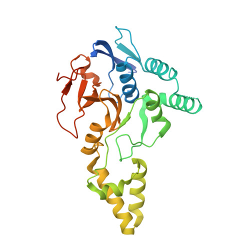
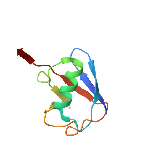
MINDY-1 Is a Member of an Evolutionarily Conserved and Structurally Distinct New Family of Deubiquitinating Enzymes.

Abdul Rehman, S.A.Kristariyanto, Y.A.Choi, S.Y.Nkosi, P.J.Weidlich, S.Labib, K.Hofmann, K.Kulathu, Y.

(2016) Mol Cell 63: 146-155

  • DOI: https://doi.org/10.1016/j.molcel.2016.05.009
  • Primary Citation Related Structures: 
    5JKN, 5JQS

  • PubMed Abstract: 

    Deubiquitinating enzymes (DUBs) remove ubiquitin (Ub) from Ub-conjugated substrates to regulate the functional outcome of ubiquitylation. Here we report the discovery of a new family of DUBs, which we have named MINDY (motif interacting with Ub-containing novel DUB family). Found in all eukaryotes, MINDY-family DUBs are highly selective at cleaving K48-linked polyUb, a signal that targets proteins for degradation. We identify the catalytic activity to be encoded within a previously unannotated domain, the crystal structure of which reveals a distinct protein fold with no homology to any of the known DUBs. The crystal structure of MINDY-1 (also known as FAM63A) in complex with propargylated Ub reveals conformational changes that realign the active site for catalysis. MINDY-1 prefers cleaving long polyUb chains and works by trimming chains from the distal end. Collectively, our results reveal a new family of DUBs that may have specialized roles in regulating proteostasis.


  • Organizational Affiliation
    • MRC Protein Phosphorylation & Ubiquitylation Unit, School of Life Sciences, University of Dundee, Dow Street, Dundee DD1 5EH, UK.

Macromolecule Content 

  • Total Structure Weight: 41.08 kDa 
  • Atom Count: 3,157 
  • Modeled Residue Count: 326 
  • Deposited Residue Count: 365 
  • Unique protein chains: 2

Macromolecules

Find similar proteins by:|  3D Structure
Entity ID: 1
MoleculeChains  Sequence LengthOrganismDetailsImage
Protein FAM63A289Homo sapiensMutation(s): 0 
Gene Names: FAM63AKIAA1390
EC: 3.4.19.12
UniProt & NIH Common Fund Data Resources
Find proteins for Q8N5J2 (Homo sapiens)
Explore Q8N5J2 
Go to UniProtKB:  Q8N5J2
PHAROS:  Q8N5J2
GTEx:  ENSG00000143409 
Entity Groups
Sequence Clusters30% Identity50% Identity70% Identity90% Identity95% Identity100% Identity
UniProt GroupQ8N5J2
Sequence Annotations
Expand
Reference Sequence
Find similar proteins by:|  3D Structure
Entity ID: 2
MoleculeChains  Sequence LengthOrganismDetailsImage
Ubiquitin-40S ribosomal protein S27aB [auth D]76Bos taurusMutation(s): 0 
Gene Names: RPS27AUBA80UBCEP1
UniProt
Find proteins for P62992 (Bos taurus)
Explore P62992 
Go to UniProtKB:  P62992
Entity Groups
Sequence Clusters30% Identity50% Identity70% Identity90% Identity95% Identity100% Identity
UniProt GroupP62992
Sequence Annotations
Expand
Reference Sequence

Small Molecules

Ligands 4 Unique
IDChains Name / Formula / InChI Key2D Diagram3D Interactions
SO4

Query on SO4


Download:Ideal Coordinates CCD File
D [auth A],
E [auth A],
H [auth D]
SULFATE ION
O4 S
QAOWNCQODCNURD-UHFFFAOYSA-L
DIO

Query on DIO


Download:Ideal Coordinates CCD File
G [auth A]1,4-DIETHYLENE DIOXIDE
C4 H8 O2
RYHBNJHYFVUHQT-UHFFFAOYSA-N
AYE

Query on AYE


Download:Ideal Coordinates CCD File
C [auth A]prop-2-en-1-amine
C3 H7 N
VVJKKWFAADXIJK-UHFFFAOYSA-N
CL

Query on CL


Download:Ideal Coordinates CCD File
F [auth A]CHLORIDE ION
Cl
VEXZGXHMUGYJMC-UHFFFAOYSA-M

Experimental Data & Validation

Experimental Data

  • Method: X-RAY DIFFRACTION
  • Resolution: 2.65 Å
  • R-Value Free:  0.231 (Depositor), 0.235 (DCC) 
  • R-Value Work:  0.205 (Depositor), 0.212 (DCC) 
  • R-Value Observed: 0.207 (Depositor) 
Space Group: P 65 2 2
Unit Cell:
Length ( Å )Angle ( ˚ )
a = 82.336α = 90
b = 82.336β = 90
c = 332.464γ = 120
Software Package:
Software NamePurpose
PHENIXrefinement
XDSdata reduction
Aimlessdata scaling
PHASERphasing

Structure Validation

View Full Validation Report


Entry History 

Deposition Data

Revision History  (Full details and data files)

  • Version 1.0: 2016-06-22
    Type: Initial release
  • Version 1.1: 2016-06-29
    Changes: Database references
  • Version 1.2: 2016-07-20
    Changes: Database references
  • Version 1.3: 2024-02-07
    Changes: Data collection, Database references, Derived calculations, Refinement description