5ILG

Crystal structure of photoreceptor dehydrogenase from Drosophila melanogaster


Experimental Data Snapshot

  • Method: X-RAY DIFFRACTION
  • Resolution: 2.40 Å
  • R-Value Free: 0.222 
  • R-Value Work: 0.171 
  • R-Value Observed: 0.174 

wwPDB Validation   3D Report Full Report


Ligand Structure Quality Assessment 


This is version 1.2 of the entry. See complete history


Literature

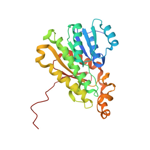
Structural Insights into the Drosophila melanogaster Retinol Dehydrogenase, a Member of the Short-Chain Dehydrogenase/Reductase Family.

Hofmann, L.Tsybovsky, Y.Alexander, N.S.Babino, D.Leung, N.Y.Montell, C.Banerjee, S.von Lintig, J.Palczewski, K.

(2016) Biochemistry 55: 6545-6557

  • DOI: https://doi.org/10.1021/acs.biochem.6b00907
  • Primary Citation of Related Structures:  
    5ILG, 5ILO

  • PubMed Abstract: 

    The 11-cis-retinylidene chromophore of visual pigments isomerizes upon interaction with a photon, initiating a downstream cascade of signaling events that ultimately lead to visual perception. 11-cis-Retinylidene is regenerated through enzymatic transformations collectively called the visual cycle. The first and rate-limiting enzymatic reaction within this cycle, i.e., the reduction of all-trans-retinal to all-trans-retinol, is catalyzed by retinol dehydrogenases. Here, we determined the structure of Drosophila melanogaster photoreceptor retinol dehydrogenase (PDH) isoform C that belongs to the short-chain dehydrogenase/reductase (SDR) family. This is the first reported structure of a SDR that possesses this biologically important activity. Two crystal structures of the same enzyme grown under different conditions revealed a novel conformational change of the NAD + cofactor, likely representing a change during catalysis. Amide hydrogen-deuterium exchange of PDH demonstrated changes in the structure of the enzyme upon dinucleotide binding. In D. melanogaster, loss of PDH activity leads to photoreceptor degeneration that can be partially rescued by transgenic expression of human RDH12. Based on the structure of PDH, we analyzed mutations causing Leber congenital amaurosis 13 in a homology model of human RDH12 to obtain insights into the molecular basis of RDH12 disease-causing mutations.


  • Organizational Affiliation

    Department of Pharmacology and Cleveland Center for Membrane and Structural Biology, School of Medicine, Case Western Reserve University , 10900 Euclid Avenue, Cleveland, Ohio 44106, United States.


Macromolecules
Find similar proteins by:  (by identity cutoff)  |  3D Structure
Entity ID: 1
MoleculeChains Sequence LengthOrganismDetailsImage
Photoreceptor dehydrogenase, isoform C
A, B
271Drosophila melanogasterMutation(s): 0 
Gene Names: PdhpcdrCG4899Dmel_CG4899
EC: 1.1.1.1 (PDB Primary Data), 1.1.1.105 (PDB Primary Data)
UniProt
Find proteins for Q9VV42 (Drosophila melanogaster)
Explore Q9VV42 
Go to UniProtKB:  Q9VV42
Entity Groups  
Sequence Clusters30% Identity50% Identity70% Identity90% Identity95% Identity100% Identity
UniProt GroupQ9VV42
Sequence Annotations
Expand
  • Reference Sequence
Small Molecules
Ligands 4 Unique
IDChains Name / Formula / InChI Key2D Diagram3D Interactions
NAD
Query on NAD

Download Ideal Coordinates CCD File 
D [auth A],
J [auth B]
NICOTINAMIDE-ADENINE-DINUCLEOTIDE
C21 H27 N7 O14 P2
BAWFJGJZGIEFAR-NNYOXOHSSA-N
IPH
Query on IPH

Download Ideal Coordinates CCD File 
E [auth A],
K [auth B]
PHENOL
C6 H6 O
ISWSIDIOOBJBQZ-UHFFFAOYSA-N
EDO
Query on EDO

Download Ideal Coordinates CCD File 
F [auth A],
G [auth A],
H [auth A]
1,2-ETHANEDIOL
C2 H6 O2
LYCAIKOWRPUZTN-UHFFFAOYSA-N
MG
Query on MG

Download Ideal Coordinates CCD File 
C [auth A],
I [auth B]
MAGNESIUM ION
Mg
JLVVSXFLKOJNIY-UHFFFAOYSA-N
Experimental Data & Validation

Experimental Data

  • Method: X-RAY DIFFRACTION
  • Resolution: 2.40 Å
  • R-Value Free: 0.222 
  • R-Value Work: 0.171 
  • R-Value Observed: 0.174 
  • Space Group: P 2 21 21
Unit Cell:
Length ( Å )Angle ( ˚ )
a = 44.195α = 90
b = 93.477β = 90
c = 114.674γ = 90
Software Package:
Software NamePurpose
PHENIXrefinement
XDSdata reduction
XDSdata scaling
PHASERphasing
MOLREPphasing

Structure Validation

View Full Validation Report


Ligand Structure Quality Assessment 


Entry History 

Deposition Data

Revision History  (Full details and data files)

  • Version 1.0: 2016-11-16
    Type: Initial release
  • Version 1.1: 2018-04-25
    Changes: Data collection, Database references, Derived calculations
  • Version 1.2: 2023-09-27
    Changes: Data collection, Database references, Refinement description