5IL1

Crystal structure of SAM-bound METTL3-METTL14 complex


Experimental Data Snapshot

  • Method: X-RAY DIFFRACTION
  • Resolution: 1.71 Å
  • R-Value Free: 0.207 
  • R-Value Work: 0.180 
  • R-Value Observed: 0.181 

wwPDB Validation   3D Report Full Report


Ligand Structure Quality Assessment 


This is version 1.1 of the entry. See complete history


Literature

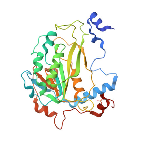
Structural basis of N6-adenosine methylation by the METTL3-METTL14 complex

Wang, X.Feng, J.Xue, Y.Guan, Z.Zhang, D.Liu, Z.Gong, Z.Wang, Q.Huang, J.Tang, C.Zou, T.Yin, P.

(2016) Nature 534: 575-578

  • DOI: https://doi.org/10.1038/nature18298
  • Primary Citation of Related Structures:  
    5IL0, 5IL1, 5IL2

  • PubMed Abstract: 

    Chemical modifications of RNA have essential roles in a vast range of cellular processes. N(6)-methyladenosine (m(6)A) is an abundant internal modification in messenger RNA and long non-coding RNA that can be dynamically added and removed by RNA methyltransferases (MTases) and demethylases, respectively. An MTase complex comprising methyltransferase-like 3 (METTL3) and methyltransferase-like 14 (METTL14) efficiently catalyses methyl group transfer. In contrast to the well-studied DNA MTase, the exact roles of these two RNA MTases in the complex remain to be elucidated. Here we report the crystal structures of the METTL3-METTL14 heterodimer with MTase domains in the ligand-free, S-adenosyl methionine (AdoMet)-bound and S-adenosyl homocysteine (AdoHcy)-bound states, with resolutions of 1.9, 1.71 and 1.61 Å, respectively. Both METTL3 and METTL14 adopt a class I MTase fold and they interact with each other via an extensive hydrogen bonding network, generating a positively charged groove. Notably, AdoMet was observed in only the METTL3 pocket and not in METTL14. Combined with biochemical analysis, these results suggest that in the m(6)A MTase complex, METTL3 primarily functions as the catalytic core, while METTL14 serves as an RNA-binding platform, reminiscent of the target recognition domain of DNA N(6)-adenine MTase. This structural information provides an important framework for the functional investigation of m(6)A.


  • Organizational Affiliation

    National Key Laboratory of Crop Genetic Improvement and National Centre of Plant Gene Research, Huazhong Agricultural University, Wuhan 430070, China.


Macromolecules
Find similar proteins by:  (by identity cutoff)  |  3D Structure
Entity ID: 1
MoleculeChains Sequence LengthOrganismDetailsImage
METTL3212Homo sapiensMutation(s): 0 
Gene Names: METTL3
EC: 2.1.1.62
UniProt & NIH Common Fund Data Resources
Find proteins for Q86U44 (Homo sapiens)
Explore Q86U44 
Go to UniProtKB:  Q86U44
PHAROS:  Q86U44
GTEx:  ENSG00000165819 
Entity Groups  
Sequence Clusters30% Identity50% Identity70% Identity90% Identity95% Identity100% Identity
UniProt GroupQ86U44
Sequence Annotations
Expand
  • Reference Sequence
Find similar proteins by:  (by identity cutoff)  |  3D Structure
Entity ID: 2
MoleculeChains Sequence LengthOrganismDetailsImage
METTL14300Homo sapiensMutation(s): 0 
Gene Names: METTL14
EC: 2.1.1.62
UniProt & NIH Common Fund Data Resources
Find proteins for Q9HCE5 (Homo sapiens)
Explore Q9HCE5 
Go to UniProtKB:  Q9HCE5
PHAROS:  Q9HCE5
GTEx:  ENSG00000145388 
Entity Groups  
Sequence Clusters30% Identity50% Identity70% Identity90% Identity95% Identity100% Identity
UniProt GroupQ9HCE5
Sequence Annotations
Expand
  • Reference Sequence
Small Molecules
Modified Residues  1 Unique
IDChains TypeFormula2D DiagramParent
SEP
Query on SEP
B
L-PEPTIDE LINKINGC3 H8 N O6 PSER
Binding Affinity Annotations 
IDSourceBinding Affinity
SAM Binding MOAD:  5IL1 Kd: 1500 (nM) from 1 assay(s)
Experimental Data & Validation

Experimental Data

  • Method: X-RAY DIFFRACTION
  • Resolution: 1.71 Å
  • R-Value Free: 0.207 
  • R-Value Work: 0.180 
  • R-Value Observed: 0.181 
  • Space Group: P 41 21 2
Unit Cell:
Length ( Å )Angle ( ˚ )
a = 102.344α = 90
b = 102.344β = 90
c = 116.72γ = 90
Software Package:
Software NamePurpose
PHENIXrefinement
XDSdata processing
Aimlessdata scaling

Structure Validation

View Full Validation Report


Ligand Structure Quality Assessment 


Entry History 

Deposition Data

Revision History  (Full details and data files)

  • Version 1.0: 2016-05-25
    Type: Initial release
  • Version 1.1: 2016-06-29
    Changes: Database references