5IBW | pdb_00005ibw

Complex of MlcC bound to the tandem IQ motif of MyoC


Experimental Data Snapshot

  • Method: X-RAY DIFFRACTION
  • Resolution: 1.90 Å
  • R-Value Free: 
    0.190 (Depositor), 0.190 (DCC) 
  • R-Value Work: 
    0.140 (Depositor), 0.145 (DCC) 

Starting Model: experimental
View more details

wwPDB Validation 3D Report Full Report

Validation slider image for 5IBW

This is version 1.5 of the entry. See complete history

Literature

Structure of the Single-lobe Myosin Light Chain C in Complex with the Light Chain-binding Domains of Myosin-1C Provides Insights into Divergent IQ Motif Recognition.

Langelaan, D.N.Liburd, J.Yang, Y.Miller, E.Chitayat, S.Crawley, S.W.Cote, G.P.Smith, S.P.

(2016) J Biological Chem 291: 19607-19617

  • DOI: https://doi.org/10.1074/jbc.M116.746313
  • Primary Citation Related Structures: 
    2M8U, 5IBW

  • PubMed Abstract: 

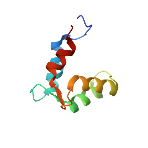
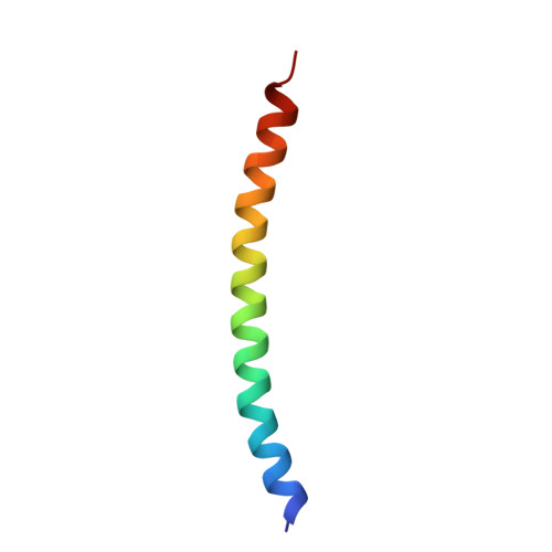
    Myosin light chains are key regulators of class 1 myosins and typically comprise two domains, with calmodulin being the archetypal example. They bind IQ motifs within the myosin neck region and amplify conformational changes in the motor domain. A single lobe light chain, myosin light chain C (MlcC), was recently identified and shown to specifically bind to two sequentially divergent IQ motifs of the Dictyostelium myosin-1C. To provide a molecular basis of this interaction, the structures of apo-MlcC and a 2:1 MlcC·myosin-1C neck complex were determined. The two non-functional EF-hand motifs of MlcC pack together to form a globular four-helix bundle that opens up to expose a central hydrophobic groove, which interacts with the N-terminal portion of the divergent IQ1 and IQ2 motifs. The N- and C-terminal regions of MlcC make critical contacts that contribute to its specific interactions with the myosin-1C divergent IQ motifs, which are contacts that deviate from the traditional mode of calmodulin-IQ recognition.


  • Organizational Affiliation
    • From the Department of Biomedical and Molecular Sciences, Queen's University, Kingston, Ontario K7L 3N6, Canada.

Macromolecule Content 

  • Total Structure Weight: 23.32 kDa 
  • Atom Count: 1,845 
  • Modeled Residue Count: 192 
  • Deposited Residue Count: 197 
  • Unique protein chains: 2

Macromolecules

Find similar proteins by:|  3D Structure
Entity ID: 1
MoleculeChains  Sequence LengthOrganismDetailsImage
Calcium-binding EF-hand domain-containing protein
A, B
77Dictyostelium discoideumMutation(s): 0 
Gene Names: DDB_G0289563
UniProt
Find proteins for Q54HC2 (Dictyostelium discoideum)
Explore Q54HC2 
Go to UniProtKB:  Q54HC2
Entity Groups
Sequence Clusters30% Identity50% Identity70% Identity90% Identity95% Identity100% Identity
UniProt GroupQ54HC2
Sequence Annotations
Expand
Reference Sequence
Find similar proteins by:|  3D Structure
Entity ID: 2
MoleculeChains  Sequence LengthOrganismDetailsImage
Myosin IC heavy chain43Dictyostelium discoideumMutation(s): 0 
Gene Names: myoCdmiCDDB_G0276617
UniProt
Find proteins for P42522 (Dictyostelium discoideum)
Explore P42522 
Go to UniProtKB:  P42522
Entity Groups
Sequence Clusters30% Identity50% Identity70% Identity90% Identity95% Identity100% Identity
UniProt GroupP42522
Sequence Annotations
Expand
Reference Sequence

Small Molecules

Ligands 1 Unique
IDChains Name / Formula / InChI Key2D Diagram3D Interactions
NA

Query on NA


Download:Ideal Coordinates CCD File
D [auth A]SODIUM ION
Na
FKNQFGJONOIPTF-UHFFFAOYSA-N

Experimental Data & Validation

Experimental Data

  • Method: X-RAY DIFFRACTION
  • Resolution: 1.90 Å
  • R-Value Free:  0.190 (Depositor), 0.190 (DCC) 
  • R-Value Work:  0.140 (Depositor), 0.145 (DCC) 
Space Group: P 1
Unit Cell:
Length ( Å )Angle ( ˚ )
a = 28.68α = 75.79
b = 31.41β = 84
c = 56.41γ = 67.06
Software Package:
Software NamePurpose
XSCALEdata scaling
PHENIXrefinement
PDB_EXTRACTdata extraction
XDSdata reduction
PHASERphasing

Structure Validation

View Full Validation Report


Entry History 

& Funding Information

Deposition Data


Funding OrganizationLocationGrant Number
Natural Sciences and Engineering Research Council (NSERC, Canada)Canada--

Revision History  (Full details and data files)

  • Version 1.0: 2016-08-03
    Type: Initial release
  • Version 1.1: 2016-08-10
    Changes: Database references
  • Version 1.2: 2016-09-21
    Changes: Database references
  • Version 1.3: 2017-09-13
    Changes: Author supporting evidence, Database references, Derived calculations
  • Version 1.4: 2020-01-08
    Changes: Author supporting evidence
  • Version 1.5: 2023-09-27
    Changes: Data collection, Database references, Refinement description