5H4R | pdb_00005h4r

the complex of Glycoside Hydrolase 5 Lichenase from Caldicellulosiruptor sp. F32 E188Q mutant and cellotetraose


Experimental Data Snapshot

  • Method: X-RAY DIFFRACTION
  • Resolution: 1.70 Å
  • R-Value Free: 
    0.170 (Depositor), 0.173 (DCC) 
  • R-Value Work: 
    0.141 (Depositor), 0.144 (DCC) 
  • R-Value Observed: 
    0.143 (Depositor) 

Starting Model: experimental
View more details

wwPDB Validation 3D Report Full Report

Validation slider image for 5H4R

This is version 2.1 of the entry. See complete history

Literature

Structural insights into the substrate specificity of a glycoside hydrolase family 5 lichenase from Caldicellulosiruptor sp. F32

Meng, D.D.Liu, X.Dong, S.Wang, Y.F.Ma, X.Q.Zhou, H.Wang, X.Yao, L.S.Feng, Y.Li, F.L.

(2017) Biochem J 474: 3373-3389

  • DOI: https://doi.org/10.1042/BCJ20170328
  • Primary Citation Related Structures: 
    4X0V, 5H4R

  • PubMed Abstract: 

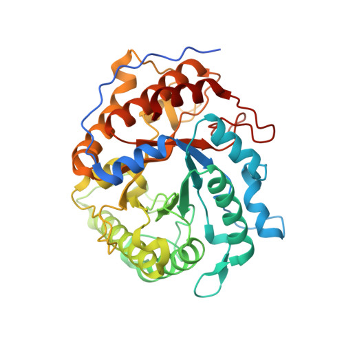
    Glycoside hydrolase (GH) family 5 is one of the largest GH families with various GH activities including lichenase, but the structural basis of the GH5 lichenase activity is still unknown. A novel thermostable lichenase F32EG5 belonging to GH5 was identified from an extremely thermophilic bacterium Caldicellulosiruptor sp. F32. F32EG5 is a bi-functional cellulose and a lichenan-degrading enzyme, and exhibited a high activity on β-1,3-1,4-glucan but side activity on cellulose. Thin-layer chromatography and NMR analyses indicated that F32EG5 cleaved the β-1,4 linkage or the β-1,3 linkage while a 4- O -substitued glucose residue linked to a glucose residue through a β-1,3 linkage, which is completely different from extensively studied GH16 lichenase that catalyses strict endo-hydrolysis of the β-1,4-glycosidic linkage adjacent to a 3- O -substitued glucose residue in the mixed-linked β-glucans. The crystal structure of F32EG5 was determined to 2.8 Å resolution, and the crystal structure of the complex of F32EG5 E193Q mutant and cellotetraose was determined to 1.7 Å resolution, which revealed that the exit subsites of substrate-binding sites contribute to both thermostability and substrate specificity of F32EG5. The sugar chain showed a sharp bend in the complex structure, suggesting that a substrate cleft fitting to the bent sugar chains in lichenan is a common feature of GH5 lichenases. The mechanism of thermostability and substrate selectivity of F32EG5 was further demonstrated by molecular dynamics simulation and site-directed mutagenesis. These results provide biochemical and structural insights into thermostability and substrate selectivity of GH5 lichenases, which have potential in industrial processes.


  • Organizational Affiliation
    • Key Laboratory of Biofuels, Qingdao Institute of Bioenergy and Bioprocess Technology, Chinese Academy of Sciences, Qingdao 266101, China.

Macromolecule Content 

  • Total Structure Weight: 46.64 kDa 
  • Atom Count: 3,583 
  • Modeled Residue Count: 375 
  • Deposited Residue Count: 396 
  • Unique protein chains: 1

Macromolecules

Find similar proteins by:|  3D Structure
Entity ID: 1
MoleculeChains  Sequence LengthOrganismDetailsImage
Beta-1,3-1,4-glucanase396Caldicellulosiruptor sp. F32Mutation(s): 1 
UniProt
Find proteins for A0ACD6B8P5 (Caldicellulosiruptor sp. F32)
Explore A0ACD6B8P5 
Go to UniProtKB:  A0ACD6B8P5
Entity Groups
Sequence Clusters30% Identity50% Identity70% Identity90% Identity95% Identity100% Identity
UniProt GroupA0ACD6B8P5
Sequence Annotations
Expand
Reference Sequence

Oligosaccharides

Help  
Entity ID: 2
MoleculeChains Length2D Diagram GlycosylationD Interactions
beta-D-glucopyranose-(1-4)-beta-D-glucopyranose-(1-4)-beta-D-glucopyranose-(1-4)-beta-D-glucopyranose
B
4N/A
Glycosylation Resources
GlyTouCan: G00025MO
GlyCosmos: G00025MO
GlyGen: G00025MO

Small Molecules

Ligands 1 Unique
IDChains Name / Formula / InChI Key2D Diagram3D Interactions
GOL

Query on GOL


Download:Ideal Coordinates CCD File
C [auth A]GLYCEROL
C3 H8 O3
PEDCQBHIVMGVHV-UHFFFAOYSA-N

Biologically Interesting Molecules (External Reference) 

1 Unique

Experimental Data & Validation

Experimental Data

  • Method: X-RAY DIFFRACTION
  • Resolution: 1.70 Å
  • R-Value Free:  0.170 (Depositor), 0.173 (DCC) 
  • R-Value Work:  0.141 (Depositor), 0.144 (DCC) 
  • R-Value Observed: 0.143 (Depositor) 
Space Group: P 21 21 21
Unit Cell:
Length ( Å )Angle ( ˚ )
a = 62.581α = 90
b = 76.315β = 90
c = 95.611γ = 90
Software Package:
Software NamePurpose
PHENIXrefinement
Aimlessdata scaling
PHASERdata processing
PDB_EXTRACTdata extraction
PHASERphasing

Structure Validation

View Full Validation Report


Entry History 

Deposition Data

Revision History  (Full details and data files)

  • Version 1.0: 2017-09-13
    Type: Initial release
  • Version 1.1: 2017-10-04
    Changes: Database references
  • Version 2.0: 2020-07-29
    Type: Remediation
    Reason: Carbohydrate remediation
    Changes: Advisory, Atomic model, Data collection, Derived calculations, Non-polymer description, Structure summary
  • Version 2.1: 2023-11-08
    Changes: Data collection, Database references, Refinement description, Structure summary