5GW7

Crystal structure of the glycosynthase mutant E727A of Escherichia coli GH63 glycosidase in complex with glucose and lactose


Experimental Data Snapshot

  • Method: X-RAY DIFFRACTION
  • Resolution: 2.20 Å
  • R-Value Free: 
    0.208 (Depositor), 0.210 (DCC) 
  • R-Value Work: 
    0.154 (Depositor), 0.160 (DCC) 
  • R-Value Observed: 
    0.157 (Depositor) 

Starting Model: experimental
View more details

wwPDB Validation   3D Report Full Report


Ligand Structure Quality Assessment 

Created with Raphaël 2.3.0Worse 01 BetterLigand structure goodness of fit to experimental dataBest fitted BGCClick on this verticalbar to view details

This is version 2.2 of the entry. See complete history


Literature

Crystal structure of the enzyme-product complex reveals sugar ring distortion during catalysis by family 63 inverting alpha-glycosidase

Miyazaki, T.Nishikawa, A.Tonozuka, T.

(2016) J Struct Biol 196: 479-486

  • DOI: https://doi.org/10.1016/j.jsb.2016.09.015
  • Primary Citation of Related Structures:  
    5CA3, 5GW7

  • PubMed Abstract: 

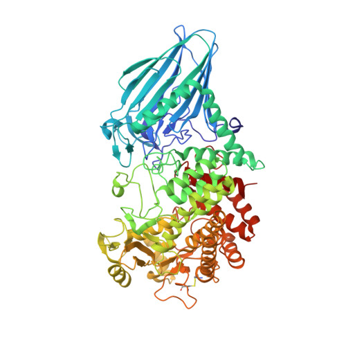
    Glycoside hydrolases are divided into two groups, known as inverting and retaining enzymes, based on their hydrolytic mechanisms. Glycoside hydrolase family 63 (GH63) is composed of inverting α-glycosidases, which act mainly on α-glucosides. We previously found that Escherichia coli GH63 enzyme, YgjK, can hydrolyze 2-O-α-d-glucosyl-d-galactose. Two constructed glycosynthase mutants, D324N and E727A, which catalyze the transfer of a β-glucosyl fluoride donor to galactose, lactose, and melibiose. Here, we determined the crystal structures of D324N and E727A soaked with a mixture of glucose and lactose at 1.8- and 2.1-Å resolutions, respectively. Because glucose and lactose molecules are found at the active sites in both structures, it is possible that these structures mimic the enzyme-product complex of YgjK. A glucose molecule found at subsite -1 in both structures adopts an unusual 1 S 3 skew-boat conformation. Comparison between these structures and the previously determined enzyme-substrate complex structure reveals that the glucose pyranose ring might be distorted immediately after nucleophilic attack by a water molecule. These structures represent the first enzyme-product complex for the GH63 family, as well as the structurally-related glycosidases, and it may provide insight into the catalytic mechanism of these enzymes.


  • Organizational Affiliation

    Research Institute of Green Science and Technology, Shizuoka University, 836 Ohya, Suruga-ku, Shizuoka 422-8529 Japan; Department of Applied Biological Science, Tokyo University of Agriculture and Technology, 3-5-8 Saiwai-cho, Fuchu, Tokyo 183-8509, Japan.


Macromolecules
Find similar proteins by:  (by identity cutoff)  |  3D Structure
Entity ID: 1
MoleculeChains Sequence LengthOrganismDetailsImage
Glucosidase YgjK
A, B
760Escherichia coli K-12Mutation(s): 1 
Gene Names: ygjK
EC: 3.2.1
UniProt
Find proteins for P42592 (Escherichia coli (strain K12))
Explore P42592 
Go to UniProtKB:  P42592
Entity Groups  
Sequence Clusters30% Identity50% Identity70% Identity90% Identity95% Identity100% Identity
UniProt GroupP42592
Sequence Annotations
Expand
  • Reference Sequence
Oligosaccharides

Help

Entity ID: 2
MoleculeChains Length2D Diagram Glycosylation3D Interactions
beta-D-galactopyranose-(1-4)-alpha-D-glucopyranose
C, D
2N/A
Glycosylation Resources
GlyTouCan:  G88362QR
GlyCosmos:  G88362QR
GlyGen:  G88362QR
Biologically Interesting Molecules (External Reference) 1 Unique
Experimental Data & Validation

Experimental Data

  • Method: X-RAY DIFFRACTION
  • Resolution: 2.20 Å
  • R-Value Free:  0.208 (Depositor), 0.210 (DCC) 
  • R-Value Work:  0.154 (Depositor), 0.160 (DCC) 
  • R-Value Observed: 0.157 (Depositor) 
Space Group: P 1 21 1
Unit Cell:
Length ( Å )Angle ( ˚ )
a = 56.74α = 90
b = 137.069β = 100
c = 81.599γ = 90
Software Package:
Software NamePurpose
REFMACrefinement
HKL-2000data scaling
MOLREPphasing
Cootmodel building

Structure Validation

View Full Validation Report


Ligand Structure Quality Assessment 

Created with Raphaël 2.3.0Worse 01 BetterLigand structure goodness of fit to experimental dataBest fitted BGCClick on this verticalbar to view details

Entry History 

Deposition Data

Revision History  (Full details and data files)

  • Version 1.0: 2016-10-12
    Type: Initial release
  • Version 1.1: 2016-12-07
    Changes: Database references
  • Version 1.2: 2020-02-26
    Changes: Data collection, Database references
  • Version 2.0: 2020-07-29
    Type: Remediation
    Reason: Carbohydrate remediation
    Changes: Atomic model, Data collection, Derived calculations, Non-polymer description, Structure summary
  • Version 2.1: 2023-11-08
    Changes: Data collection, Database references, Refinement description, Structure summary
  • Version 2.2: 2024-11-13
    Changes: Structure summary