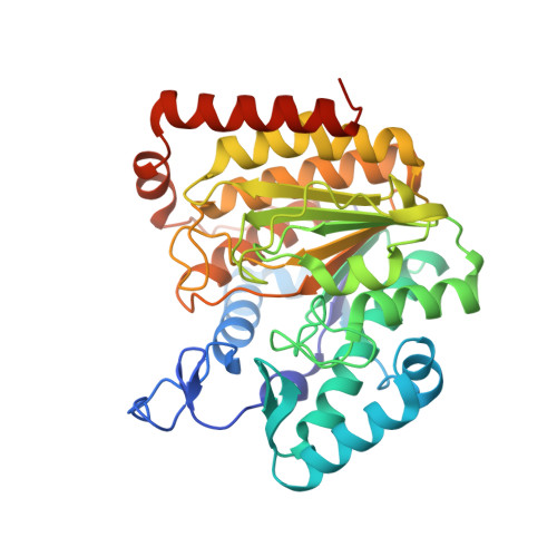
5G10

Pseudomonas aeruginosa HDAH bound to 9,9,9 trifluoro-8,8-dihydroy-N-phenylnonanamide


Experimental Data Snapshot

  • Method: X-RAY DIFFRACTION
  • Resolution: 1.71 Å
  • R-Value Free: 0.194 
  • R-Value Work: 0.158 
  • R-Value Observed: 0.160 

Starting Model: experimental
View more details

wwPDB Validation   3D Report Full Report


Ligand Structure Quality Assessment 


This is version 1.2 of the entry. See complete history


Literature

Crystal Structure of a Histone Deacetylase Homologue from Pseudomonas aeruginosa.

Kramer, A.Wagner, T.Yildiz, O.Meyer-Almes, F.J.

(2016) Biochemistry 55: 6858-6868

  • DOI: https://doi.org/10.1021/acs.biochem.6b00613
  • Primary Citation of Related Structures:  
    5G0X, 5G0Y, 5G10, 5G11, 5G12, 5G13

  • PubMed Abstract: 

    Despite the recently growing interest in the acetylation of lysine residues by prokaryotic enzymes, the underlying biological function is still not well understood. Deacetylation is accomplished by proteins that belong to the histone deacetylase (HDAC) superfamily. In this report, we present the first crystal structure of PA3774, a histone deacetylase homologue from the human pathogen Pseudomonas aeruginosa that shares a high degree of homology with class IIb HDACs. We determined the crystal structure of the ligand-free enzyme and protein-ligand complexes with a trifluoromethylketone inhibitor and the reaction product acetate. Moreover, we produced loss of function mutants and determined the structure of the inhibitor-free PA3774 H143A mutant, the inhibitor-free PA3774 Y313F mutant, and the PA3774 Y313F mutant in complex with the highly selective hydroxamate inhibitor PFSAHA. The overall structure reveals that the exceptionally long L1 loop mediates the formation of a tetramer composed of two "head-to-head" dimers. The distinctive dimer interface significantly confines the entrance area of the active site, suggesting a crucial role for substrate recognition and selectivity.


  • Organizational Affiliation

    University of Applied Sciences , Department of Chemical Engineering and Biotechnology, 64295 Darmstadt, Germany.


Macromolecules
Find similar proteins by:  (by identity cutoff)  |  3D Structure
Entity ID: 1
MoleculeChains Sequence LengthOrganismDetailsImage
HDAH
A, B
379Pseudomonas aeruginosa PAO1Mutation(s): 0 
EC: 3.5.1
UniProt
Find proteins for Q9HXM1 (Pseudomonas aeruginosa (strain ATCC 15692 / DSM 22644 / CIP 104116 / JCM 14847 / LMG 12228 / 1C / PRS 101 / PAO1))
Explore Q9HXM1 
Go to UniProtKB:  Q9HXM1
Entity Groups  
Sequence Clusters30% Identity50% Identity70% Identity90% Identity95% Identity100% Identity
UniProt GroupQ9HXM1
Sequence Annotations
Expand
  • Reference Sequence
Small Molecules
Ligands 3 Unique
IDChains Name / Formula / InChI Key2D Diagram3D Interactions
6DK
Query on 6DK

Download Ideal Coordinates CCD File 
F [auth A],
J [auth B]
9,9,9-tris(fluoranyl)-8,8-bis(oxidanyl)-~{N}-phenyl-nonanamide
C15 H20 F3 N O3
DOVGAPNVSPZBAT-UHFFFAOYSA-N
ZN
Query on ZN

Download Ideal Coordinates CCD File 
C [auth A],
G [auth B]
ZINC ION
Zn
PTFCDOFLOPIGGS-UHFFFAOYSA-N
K
Query on K

Download Ideal Coordinates CCD File 
D [auth A],
E [auth A],
H [auth B],
I [auth B]
POTASSIUM ION
K
NPYPAHLBTDXSSS-UHFFFAOYSA-N
Experimental Data & Validation

Experimental Data

  • Method: X-RAY DIFFRACTION
  • Resolution: 1.71 Å
  • R-Value Free: 0.194 
  • R-Value Work: 0.158 
  • R-Value Observed: 0.160 
  • Space Group: P 41 21 2
Unit Cell:
Length ( Å )Angle ( ˚ )
a = 81.74α = 90
b = 81.74β = 90
c = 205.21γ = 90
Software Package:
Software NamePurpose
REFMACrefinement
MOSFLMdata reduction
Aimlessdata scaling
MOLREPphasing

Structure Validation

View Full Validation Report


Ligand Structure Quality Assessment 


Entry History 

Deposition Data

Revision History  (Full details and data files)

  • Version 1.0: 2016-11-30
    Type: Initial release
  • Version 1.1: 2017-03-01
    Changes: Database references
  • Version 1.2: 2024-01-10
    Changes: Data collection, Database references, Derived calculations, Other, Refinement description