5DOF | pdb_00005dof

Crystal structure of Tetrahymena p19


Experimental Data Snapshot

  • Method: X-RAY DIFFRACTION
  • Resolution: 1.70 Å
  • R-Value Free: 
    0.220 (Depositor), 0.219 (DCC) 
  • R-Value Work: 
    0.177 (Depositor), 0.176 (DCC) 
  • R-Value Observed: 
    0.179 (Depositor) 

wwPDB Validation 3D Report Full Report

Validation slider image for 5DOF

This is version 1.2 of the entry. See complete history

Literature

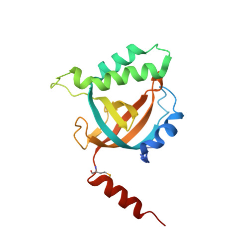
The Tetrahymena telomerase p75-p45-p19 subcomplex is a unique CST complex.

Wan, B.Tang, T.Upton, H.Shuai, J.Zhou, Y.Li, S.Chen, J.Brunzelle, J.S.Zeng, Z.Collins, K.Wu, J.Lei, M.

(2015) Nat Struct Mol Biol 22: 1023-1026

  • DOI: https://doi.org/10.1038/nsmb.3126
  • Primary Citation Related Structures: 
    5DOF, 5DOI, 5DOK

  • PubMed Abstract: 

    Tetrahymena telomerase holoenzyme subunits p75, p45 and p19 form a subcomplex (7-4-1) peripheral to the catalytic core. We report structures of p45 and p19 and reveal them as the Stn1 and Ten1 subunits of the CST complex, which stimulates telomerase complementary-strand synthesis. 7-4-1 binds telomeric single-stranded DNA, and mutant p19 overexpression causes telomere 3'-overhang elongation. We propose that telomerase-tethered Tetrahymena CST coordinates telomere G-strand and C-strand synthesis.


  • Organizational Affiliation
    • National Center for Protein Science Shanghai, State Key Laboratory of Molecular Biology, Institute of Biochemistry and Cell Biology, Shanghai Institutes for Biological Sciences, Chinese Academy of Sciences, Shanghai, China.

Macromolecule Content 

  • Total Structure Weight: 77.65 kDa 
  • Atom Count: 5,735 
  • Modeled Residue Count: 612 
  • Deposited Residue Count: 652 
  • Unique protein chains: 1

Macromolecules

Find similar proteins by:|  3D Structure
Entity ID: 1
MoleculeChains  Sequence LengthOrganismDetailsImage
Telomerase-associated protein 19
A, B, C, D
163Tetrahymena thermophilaMutation(s): 0 
Gene Names: TAP19
UniProt
Find proteins for D2CVN7 (Tetrahymena thermophila (strain SB210))
Explore D2CVN7 
Go to UniProtKB:  D2CVN7
Entity Groups
Sequence Clusters30% Identity50% Identity70% Identity90% Identity95% Identity100% Identity
UniProt GroupD2CVN7
Sequence Annotations
Expand
Reference Sequence

Small Molecules

Modified Residues  1 Unique
IDChains TypeFormula2D DiagramParent
MSE
Query on MSE
A, B, C, D
L-PEPTIDE LINKINGC5 H11 N O2 SeMET

Experimental Data & Validation

Experimental Data

  • Method: X-RAY DIFFRACTION
  • Resolution: 1.70 Å
  • R-Value Free:  0.220 (Depositor), 0.219 (DCC) 
  • R-Value Work:  0.177 (Depositor), 0.176 (DCC) 
  • R-Value Observed: 0.179 (Depositor) 
Space Group: P 1 21 1
Unit Cell:
Length ( Å )Angle ( ˚ )
a = 40.732α = 90
b = 128.631β = 105.05
c = 75.328γ = 90
Software Package:
Software NamePurpose
HKL-2000data scaling
REFMACrefinement
PDB_EXTRACTdata extraction
HKL-2000data collection
Cootmodel building

Structure Validation

View Full Validation Report


Entry History 

Deposition Data

Revision History  (Full details and data files)

  • Version 1.0: 2015-11-25
    Type: Initial release
  • Version 1.1: 2015-12-23
    Changes: Database references
  • Version 1.2: 2024-10-09
    Changes: Data collection, Database references, Derived calculations, Structure summary