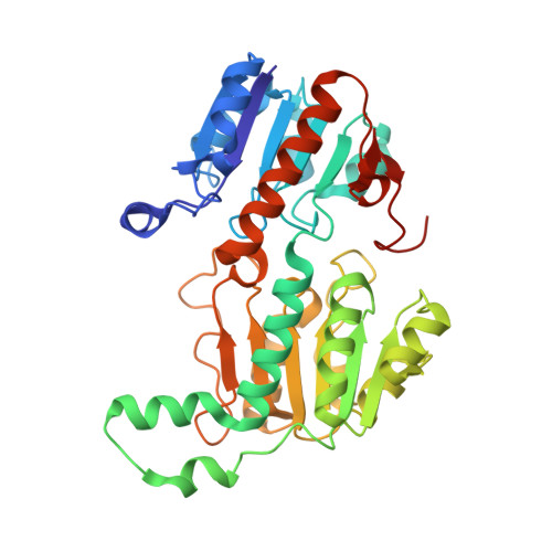
5DN9

Crystal structure of Candida boidinii formate dehydrogenase complexed with NAD+ and azide


Experimental Data Snapshot

  • Method: X-RAY DIFFRACTION
  • Resolution: 1.50 Å
  • R-Value Free: 0.169 
  • R-Value Work: 0.138 
  • R-Value Observed: 0.140 

Starting Model: experimental
View more details

wwPDB Validation   3D Report Full Report


Ligand Structure Quality Assessment 


This is version 1.4 of the entry. See complete history


Literature

Structural and Kinetic Studies of Formate Dehydrogenase from Candida boidinii.

Guo, Q.Gakhar, L.Wickersham, K.Francis, K.Vardi-Kilshtain, A.Major, D.T.Cheatum, C.M.Kohen, A.

(2016) Biochemistry 55: 2760-2771

  • DOI: https://doi.org/10.1021/acs.biochem.6b00181
  • Primary Citation of Related Structures:  
    5DN9, 5DNA

  • PubMed Abstract: 

    The structure of formate dehydrogenase from Candida boidinii (CbFDH) is of both academic and practical interests. First, this enzyme represents a unique model system for studies on the role of protein dynamics in catalysis, but so far these studies have been limited by the availability of structural information. Second, CbFDH and its mutants can be used in various industrial applications (e.g., CO2 fixation or nicotinamide recycling systems), and the lack of structural information has been a limiting factor in commercial development. Here, we report the crystallization and structural determination of both holo- and apo-CbFDH. The free-energy barrier for the catalyzed reaction was computed and indicates that this structure indeed represents a catalytically competent form of the enzyme. Complementing kinetic examinations demonstrate that the recombinant CbFDH has a well-organized reactive state. Finally, a fortuitous observation has been made: the apoenzyme crystal was obtained under cocrystallization conditions with a saturating concentration of both the cofactor (NAD(+)) and inhibitor (azide), which has a nanomolar dissociation constant. It was found that the fraction of the apoenzyme present in the solution is less than 1.7 × 10(-7) (i.e., the solution is 99.9999% holoenzyme). This is an extreme case where the crystal structure represents an insignificant fraction of the enzyme in solution, and a mechanism rationalizing this phenomenon is presented.


  • Organizational Affiliation

    Department of Chemistry, University of Iowa , Iowa City, Iowa 52242, United States.


Macromolecules
Find similar proteins by:  (by identity cutoff)  |  3D Structure
Entity ID: 1
MoleculeChains Sequence LengthOrganismDetailsImage
FDH
A, B
364[Candida] boidiniiMutation(s): 0 
Gene Names: FDH
EC: 1.2.1.2 (PDB Primary Data), 1.17.1.9 (UniProt)
UniProt
Find proteins for A0A0A1EQY0 (Candida boidinii)
Explore A0A0A1EQY0 
Go to UniProtKB:  A0A0A1EQY0
Entity Groups  
Sequence Clusters30% Identity50% Identity70% Identity90% Identity95% Identity100% Identity
UniProt GroupA0A0A1EQY0
Sequence Annotations
Expand
  • Reference Sequence
Experimental Data & Validation

Experimental Data

  • Method: X-RAY DIFFRACTION
  • Resolution: 1.50 Å
  • R-Value Free: 0.169 
  • R-Value Work: 0.138 
  • R-Value Observed: 0.140 
  • Space Group: P 1 21 1
Unit Cell:
Length ( Å )Angle ( ˚ )
a = 50.931α = 90
b = 116.617β = 106.9
c = 63.208γ = 90
Software Package:
Software NamePurpose
PHENIXrefinement
REFMACrefinement
XDSdata scaling
Cootmodel building
PHASERphasing
Blu-Icedata collection
SCALAdata scaling

Structure Validation

View Full Validation Report


Ligand Structure Quality Assessment 


Entry History & Funding Information

Deposition Data


Funding OrganizationLocationGrant Number
National Institutes of Health/National Institute of General Medical Sciences (NIH/NIGMS)United StatesGM065368
National Science Foundation (NSF, United States)United StatesCHE-1149023
United States - Israel Binational Science Foundation (BSF)United States2012340
National Institutes of Health/National Institute of General Medical Sciences (NIH/NIGMS)United StatesGM079368

Revision History  (Full details and data files)

  • Version 1.0: 2016-05-04
    Type: Initial release
  • Version 1.1: 2016-05-25
    Changes: Database references
  • Version 1.2: 2017-09-20
    Changes: Author supporting evidence, Database references, Derived calculations, Refinement description
  • Version 1.3: 2019-11-27
    Changes: Author supporting evidence
  • Version 1.4: 2023-09-27
    Changes: Data collection, Database references, Refinement description