5C87

Crystal structure of the human BRPF1 bromodomain in complex with SEED2


Experimental Data Snapshot

  • Method: X-RAY DIFFRACTION
  • Resolution: 1.55 Å
  • R-Value Free: 0.198 
  • R-Value Work: 0.182 
  • R-Value Observed: 0.183 

wwPDB Validation   3D Report Full Report


This is version 1.2 of the entry. See complete history


Literature

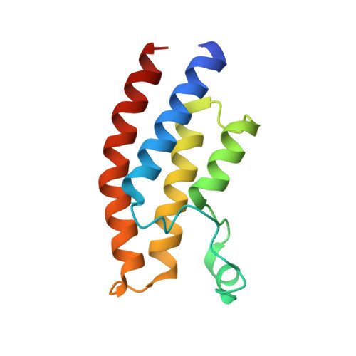
Twenty Crystal Structures of Bromodomain and PHD Finger Containing Protein 1 (BRPF1)/Ligand Complexes Reveal Conserved Binding Motifs and Rare Interactions.

Zhu, J.Caflisch, A.

(2016) J Med Chem 59: 5555-5561

  • DOI: https://doi.org/10.1021/acs.jmedchem.6b00215
  • Primary Citation of Related Structures:  
    5C7N, 5C85, 5C87, 5DY7, 5DYA, 5DYC, 5E3D, 5E3G, 5EM3, 5EPR, 5EPS, 5EQ1, 5ETB, 5ETD, 5EV9, 5EVA, 5EWC, 5EWD, 5EWH

  • PubMed Abstract: 

    BRPF1 plays a scaffolding role in transcription. We report on fragment screening by high-throughput docking to the BRPF1 bromodomain which resulted in six chemotypes with very favorable ligand efficiency (0.45-0.50 kcal/mol per non-hydrogen atom). Twenty crystal structures of BRPF1/ligand complexes show structural conservation in the acetyllysine binding site, common binding motifs, and unusual interactions (e.g., the replacement of a conserved water molecule). The structural information is useful for the design of chemical probes.


  • Organizational Affiliation

    Department of Biochemistry, University of Zürich , Winterthurerstrasse 190, CH-8057 Zürich, Switzerland.


Macromolecules
Find similar proteins by:  (by identity cutoff)  |  3D Structure
Entity ID: 1
MoleculeChains Sequence LengthOrganismDetailsImage
Peregrin116Homo sapiensMutation(s): 0 
Gene Names: BRPF1BR140
UniProt & NIH Common Fund Data Resources
Find proteins for P55201 (Homo sapiens)
Explore P55201 
Go to UniProtKB:  P55201
PHAROS:  P55201
GTEx:  ENSG00000156983 
Entity Groups  
Sequence Clusters30% Identity50% Identity70% Identity90% Identity95% Identity100% Identity
UniProt GroupP55201
Sequence Annotations
Expand
  • Reference Sequence
Small Molecules
Binding Affinity Annotations 
IDSourceBinding Affinity
4YS BindingDB:  5C87 Kd: 2.00e+5 (nM) from 1 assay(s)
Experimental Data & Validation

Experimental Data

  • Method: X-RAY DIFFRACTION
  • Resolution: 1.55 Å
  • R-Value Free: 0.198 
  • R-Value Work: 0.182 
  • R-Value Observed: 0.183 
  • Space Group: P 32 2 1
Unit Cell:
Length ( Å )Angle ( ˚ )
a = 60.559α = 90
b = 60.559β = 90
c = 63.599γ = 120
Software Package:
Software NamePurpose
PHENIXrefinement
XDSdata reduction
SCALAdata scaling
PHASERphasing

Structure Validation

View Full Validation Report


Entry History 

Deposition Data

Revision History  (Full details and data files)

  • Version 1.0: 2016-05-25
    Type: Initial release
  • Version 1.1: 2016-06-22
    Changes: Database references
  • Version 1.2: 2024-01-10
    Changes: Data collection, Database references, Refinement description