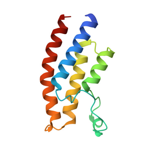
5EPS

Crystal structure of the human BRPF1 bromodomain in complex with SEED10


Experimental Data Snapshot

  • Method: X-RAY DIFFRACTION
  • Resolution: 1.47 Å
  • R-Value Free: 0.198 
  • R-Value Work: 0.188 
  • R-Value Observed: 0.189 

Starting Model: experimental
View more details

wwPDB Validation   3D Report Full Report


Ligand Structure Quality Assessment 


This is version 2.0 of the entry. See complete history


Literature

Twenty Crystal Structures of Bromodomain and PHD Finger Containing Protein 1 (BRPF1)/Ligand Complexes Reveal Conserved Binding Motifs and Rare Interactions.

Zhu, J.Caflisch, A.

(2016) J Med Chem 59: 5555-5561

  • DOI: https://doi.org/10.1021/acs.jmedchem.6b00215
  • Primary Citation of Related Structures:  
    5C7N, 5C85, 5C87, 5DY7, 5DYA, 5DYC, 5E3D, 5E3G, 5EM3, 5EPR, 5EPS, 5EQ1, 5ETB, 5ETD, 5EV9, 5EVA, 5EWC, 5EWD, 5EWH

  • PubMed Abstract: 

    BRPF1 plays a scaffolding role in transcription. We report on fragment screening by high-throughput docking to the BRPF1 bromodomain which resulted in six chemotypes with very favorable ligand efficiency (0.45-0.50 kcal/mol per non-hydrogen atom). Twenty crystal structures of BRPF1/ligand complexes show structural conservation in the acetyllysine binding site, common binding motifs, and unusual interactions (e.g., the replacement of a conserved water molecule). The structural information is useful for the design of chemical probes.


  • Organizational Affiliation

    Department of Biochemistry, University of Zürich , Winterthurerstrasse 190, CH-8057 Zürich, Switzerland.


Macromolecules
Find similar proteins by:  (by identity cutoff)  |  3D Structure
Entity ID: 1
MoleculeChains Sequence LengthOrganismDetailsImage
Peregrin116Homo sapiensMutation(s): 0 
Gene Names: BRPF1BR140
UniProt & NIH Common Fund Data Resources
Find proteins for P55201 (Homo sapiens)
Explore P55201 
Go to UniProtKB:  P55201
PHAROS:  P55201
GTEx:  ENSG00000156983 
Entity Groups  
Sequence Clusters30% Identity50% Identity70% Identity90% Identity95% Identity100% Identity
UniProt GroupP55201
Sequence Annotations
Expand
  • Reference Sequence
Small Molecules
Binding Affinity Annotations 
IDSourceBinding Affinity
5QX BindingDB:  5EPS Kd: 3.80e+4 (nM) from 1 assay(s)
Experimental Data & Validation

Experimental Data

  • Method: X-RAY DIFFRACTION
  • Resolution: 1.47 Å
  • R-Value Free: 0.198 
  • R-Value Work: 0.188 
  • R-Value Observed: 0.189 
  • Space Group: P 32 2 1
Unit Cell:
Length ( Å )Angle ( ˚ )
a = 60.63α = 90
b = 60.63β = 90
c = 62.5γ = 120
Software Package:
Software NamePurpose
PHENIXrefinement
XDSdata reduction
XSCALEdata scaling
PHASERphasing

Structure Validation

View Full Validation Report


Ligand Structure Quality Assessment 


Entry History & Funding Information

Deposition Data


Funding OrganizationLocationGrant Number
Swiss National Science FoundationSwitzerland--

Revision History  (Full details and data files)

  • Version 1.0: 2016-05-25
    Type: Initial release
  • Version 1.1: 2016-06-22
    Changes: Database references
  • Version 2.0: 2024-01-10
    Changes: Atomic model, Data collection, Database references, Refinement description