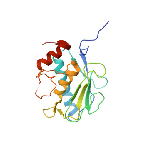
5BOY

X-RAY Co-structure of MMP-13 with ethyl 5-(1-methyl-1H-imidazol-5-yl)-1H-indole-2-Carboxylate


Experimental Data Snapshot

  • Method: X-RAY DIFFRACTION
  • Resolution: 2.03 Å
  • R-Value Free: 0.190 
  • R-Value Work: 0.160 

wwPDB Validation   3D Report Full Report


Ligand Structure Quality Assessment 


This is version 1.3 of the entry. See complete history


Literature

Fragment-based discovery of indole inhibitors of matrix metalloproteinase-13.

Taylor, S.J.Abeywardane, A.Liang, S.Muegge, I.Padyana, A.K.Xiong, Z.Hill-Drzewi, M.Farmer, B.Li, X.Collins, B.Li, J.X.Heim-Riether, A.Proudfoot, J.Zhang, Q.Goldberg, D.Zuvela-Jelaska, L.Zaher, H.Li, J.Farrow, N.A.

(2011) J Med Chem 54: 8174-8187

  • DOI: https://doi.org/10.1021/jm201129m
  • Primary Citation of Related Structures:  
    5BOT, 5BOY, 5BPA

  • PubMed Abstract: 

    Matrix metalloproteases (MMPs) play an important role in cartilage homeostasis under both normal and inflamed disease states and, thus, have become attractive targets for the treatment of arthritic diseases. Herein, we describe the identification of a potent, selective MMP-13 inhibitor, developed using fragment-based structure-guided lead identification and optimization techniques. Virtual screening methods identified a novel, indole-based MMP-13 inhibitor that bound into the S1' pocket of the protein exhibiting a novel interaction pattern hitherto not observed in MMP-13 inhibitors. X-ray crystallographic structures were used to guide the elaboration of the fragment, ultimately leading to a potent inhibitor that was >100-fold selective over nine other MMP isoforms tested.


  • Organizational Affiliation

    Boehringer Ingelheim Pharmaceuticals Inc, Ridgefield, Connecticut 06877-0368, United States. steven.taylor@boehringeringelheim.com


Macromolecules
Find similar proteins by:  (by identity cutoff)  |  3D Structure
Entity ID: 1
MoleculeChains Sequence LengthOrganismDetailsImage
Collagenase 3
A, B
171Homo sapiensMutation(s): 0 
Gene Names: MMP13
EC: 3.4.24
UniProt & NIH Common Fund Data Resources
Find proteins for P45452 (Homo sapiens)
Explore P45452 
Go to UniProtKB:  P45452
PHAROS:  P45452
GTEx:  ENSG00000137745 
Entity Groups  
Sequence Clusters30% Identity50% Identity70% Identity90% Identity95% Identity100% Identity
UniProt GroupP45452
Sequence Annotations
Expand
  • Reference Sequence
Small Molecules
Ligands 3 Unique
IDChains Name / Formula / InChI Key2D Diagram3D Interactions
4UE
Query on 4UE

Download Ideal Coordinates CCD File 
H [auth A],
N [auth B]
ethyl 5-(1-methyl-1H-imidazol-5-yl)-1H-indole-2-carboxylate
C15 H15 N3 O2
FOLNSFQIWULSJC-UHFFFAOYSA-N
ZN
Query on ZN

Download Ideal Coordinates CCD File 
C [auth A],
D [auth A],
I [auth B],
J [auth B]
ZINC ION
Zn
PTFCDOFLOPIGGS-UHFFFAOYSA-N
CA
Query on CA

Download Ideal Coordinates CCD File 
E [auth A]
F [auth A]
G [auth A]
K [auth B]
L [auth B]
E [auth A],
F [auth A],
G [auth A],
K [auth B],
L [auth B],
M [auth B]
CALCIUM ION
Ca
BHPQYMZQTOCNFJ-UHFFFAOYSA-N
Experimental Data & Validation

Experimental Data

  • Method: X-RAY DIFFRACTION
  • Resolution: 2.03 Å
  • R-Value Free: 0.190 
  • R-Value Work: 0.160 
  • Space Group: C 1 2 1
Unit Cell:
Length ( Å )Angle ( ˚ )
a = 135.079α = 90
b = 36.051β = 131.26
c = 95.908γ = 90
Software Package:
Software NamePurpose
PHENIXrefinement

Structure Validation

View Full Validation Report


Ligand Structure Quality Assessment 


Entry History 

Deposition Data

  • Released Date: 2015-06-17 
  • Deposition Author(s): Farrow, N.A.

Revision History  (Full details and data files)

  • Version 1.0: 2015-06-17
    Type: Initial release
  • Version 1.1: 2015-08-12
    Changes: Refinement description
  • Version 1.2: 2015-10-07
    Changes: Experimental preparation
  • Version 1.3: 2024-03-06
    Changes: Data collection, Database references, Derived calculations